Write your message
Volume 7, Issue 3 (August 2020)                   IJML 2020, 7(3): 197-211 | Back to browse issues page


XML Print


Download citation:
BibTeX | RIS | EndNote | Medlars | ProCite | Reference Manager | RefWorks
Send citation to:

Doctor Arastoye Marandi M, Yadegari M, Shahedi A, Pourentezari M, Anvari M, Shahrokhi Raeini A et al . Effects of Icariin on Histomorphometric Changes of Testis and Prostate Induced by Acrylamide in Mice. IJML 2020; 7 (3) :197-211
URL: http://ijml.ssu.ac.ir/article-1-347-en.html
Department of Anatomy and Cell Biology, Faculty of Medicine, Shahid Sadoughi University of Medical Sciences, Yazd, Iran
Full-Text [PDF 1022 kb]   (550 Downloads)     |   Abstract (HTML)  (924 Views)
Full-Text:   (648 Views)
Introduction
During recent decades, Acrylamide has been recognized as a risk factor for infertility. It is a white crystalline solid, which is water-and-alcohol-soluble, but insoluble in heptane and benzene. It is an odorless compound reactive that formed Acrylamide with its molecular formula C3H5NO [1]. Acrylamide monomer is widely used to manufacture polymers and copolymers for use in the mining, paper, and also the polymer industry, wastewater treatment, adhesives, laboratory gels, and oil field industries [2]. Recently, the presence of Acrylamide in a wide variety of human food, including heat-treated food products, has been reported. The formation of Acrylamide is increased with high-temperature (≤200C) processing in certain carbohydrate-rich foods such as chips, fried potatoes and bread. Besides, Acrylamide has been reported to be among carcinogen and cytotoxic material [3]. Exposure to Acrylamide can occur in workplaces or environment through air, water, land, and groundwater during production or use [4]. Additionally, Acrylamide showed reproductive toxicity, including germ cell mutagenicity and transmitted mutations. Recent reports indicated that binding of Acrylamide or glycidamide to spermatid protamine causes dominant lethal mutation of testicular germ cells and morphologic abnormalities of sperm but no significant effects on epididymal sperm count or motility [5, 6]. Some studies indicate that Acrylamide exposure can lead to testicular degeneration and damage in the testis. These changes were more severe with a more extended period of exposure effects of Acrylamide on the reproductive system of animals, including decreased sperm count, increased abnormal sperm morphology, and severe testicular damages, such as vacuolation and swelling of the round spermatid, and break of DNA during specific germ cell stages [7, 8]. Besides, animals fed with Acrylamide exhibited significant reductions in fertility, as well as sperm transport in the female reproductive tract. It is necessary to decrease Acrylamide levels in different foods, and to find ways to decrease the Acrylamide formation as much as possible during the cooking process of different foods [1].
Icariin (C33H40O15; molecular weight: 676.67), a flavonoid extracted from Herbaepimedii, is considered to be the principal active ingredient responsible for the actions of the plant. Herbaepimedii is known as an effective cure for cardiovascular diseases, osteoporosis (reduced bone mass), and tumors. Furthermore, it can improve the endocrine and immune system functioning [9, 10]. Herbaepimedii has been used in China as a remedy for the erectile dysfunction, traditionally [11]. Animal experiments showed that Icariin has an extensive range of effects on reproductive functions in male rats like improving erectile functioning in aged male rats in the way that streptozotocin-induced diabetic rats testosterone production can be increased by an accurate dose of Icariin [12]. However, an inordinate dose of Icariin can probably cause harmful effects such as tissue and organ oxidative damage [13].
There was not any published study reviewed the effect of both Acrylamide and Icariin, simultaneously. In this study, the effects of Icariin on histomorphometric changes of testis and prostate induced by Acrylamide were inspected in mice.

Materials and Methods
Chemicals
The Icariin, Acrylamide, and eosin were received from Sigma_Aldrich Chemical Company. Other chemicals such as Azocarmine B and Aniline orange G were obtained from Santa Cruz Biotechnology.
Animals
The animals were handled according to a protocol approved by the Ethical Committee of Shahid Sadoughi University of Medical Sciences, Yazd, Iran, by the code of IR.SSU.MEDICINE.REC.1397.157.
This experimental study was conducted on 32 male mice, weighing 20-35 gr, which were obtained from the animal house of Shahid Sadoughi University of Medical Sciences, Yazd, Iran. Male mice were received to acclimate under moderated humidity (40-50%), temperature (22-24°C), and light conditions (12-hr light-dark cycle) and had free access to mice food and drinking water.
Experimental design
After 7 days of adaptation to the surroundings, the mice were randomly divided into four groups (n=8): a control group, a sham group that received water, a group receiving an intraperitoneal injection of Acrylamide (10 mg/kg body weight, dissolved in water) [14] and a group receiving an intraperitoneal injection of Acrylamide (1.5 mg/kg, water solution) + Icariin (1.5 mg/kg, water solution) [13].
Tissue preparation and histological analysis of testis and prostate
On day 36, all mice were weighed and anesthetized by chloroform; heart blood samples were subsequently taken and analyzed by mouse testosterone enzyme-linked immunosorbent assay  kit for the quantitative determination of testosterone in mouse serum for each animal. The testosterone kit was purchased from Sigma Company. An analytic scale weighted the left testis and prostate of each mouse with a precision of 0.001gr. The testes and prostate were fixed in Bouin's and formalin solution, respectively, at room temperature for at least 24 h. It will slowly penetrate the tissue causing chemical and physical changes that harden and preserve the tissue and protect it against subsequent processing steps [
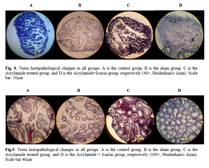
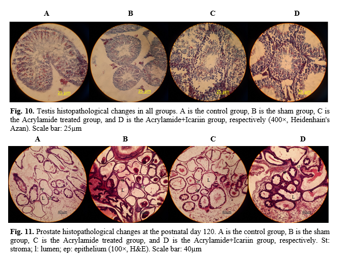
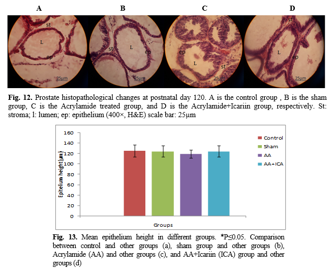
15]. The next steps were dehydration, clearing, and wax infiltration. The tissues were processed for paraffin blocking out and sectioning. Then, 5-µm serial microscopic sections with a certain distance were prepared, were taken from each testis, and serial sections were stained with hematoxylin and eosin and Heidenhain's Azan. The stained sections were mounted and examined under light microscopy (Olympus Japan) with the magnification of 40×, 100×, 400× to evaluate spermatogenesis and histopathological changes of the prostate.
The maturity of the seminiferous epithelium was marked by using a modified Johnsen's score, which was used to categorize the spermatogenesis. This method applies a score of 1-10 for each tubule cross-section that was examined. The germinal epithelium of at least 50-60 tubules was assessed for each testis, and the mean Johnsen's score per mice was calculated [16-18].
A grade from 1 to 10 was given to each tubule cross-section according to the following criteria: 10 = full spermatogenesis and perfect tubules; 9= many late spermatids and disorganized spermatogenesis; 8= only less than five spermatozoa per tubule, few late spermatids were present; 7= no spermatozoa, no late spermatids but many early spermatids present; 6= only a few early spermatids present, the arrest of spermatogenesis at the spermatid stage, disturbance of spermatid differentiation; 5= no spermatozoa or spermatids but many spermatocytes were present; 4= only a few spermatocytes were present, the arrest of spermatogenesis at the primary spermatocyte stage; 3= only spermatogonia was present; 2= no germinal cells but only sertoli cells were present; 1= tubular sclerosis, no germ cells and no sertoli cells were present.
Heidenhain's Azan staining
A technique used in stereological studies on the testis, using azocarmine B followed by aniline blue to stain nuclei red, cytoplasm pale pink, and the connective tissue blue [19, 20]. This method differentiates the cells. Thus, cells can be easier investigated and counted.
Heidenhain's Azan Technique
The procedure is as follows:
1) Samples were stained in 1% Azocarmine B solution for about 15 minutes at room temperature. 2) Rinsed in distilled water. 3) Sections were stained in aniline for 2 seconds. 4) The samples were treated with glacial acetic acid for 5 seconds. 5) They were in phosphotungstic acid for 2-3 minutes. 6) They have transferred to an aniline blue-orange G-acetic solution for 5 minutes. 7) They were rinsed with distilled water.
Stereological analysis of testis
The volume of the testis (mm3)
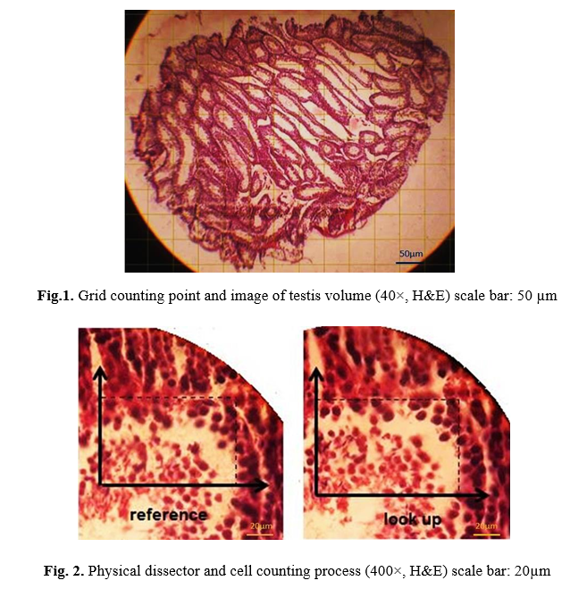
To estimate the total size and volume of the testes in mice, Cavalieri's principle was used because Cavalieri principle runs free from the shape of a solid object. Following histological procedures, the paraffin-embedded testis specimens were sectioned at 5 mm by the microtome with systematic random sampling. Serial sections, with a specific thickness, and a specific distance, have been prepared (5 µm) consecutively from each testis. A point counting grid with a 100 μm distance between two points was used to estimate the area of each section of the testis, and it was randomly located over images (since the point is lacking dimension, a grid with a network of crosses (+) was used (Fig. 1).
Then, hitting points on the surface of the biopsy were counted, and the total volume of the testis was calculated using the following formula:
V= t × a/p × ∑P/m2
V is the total volume of testis, t is defined as means section thickness, a/p; area per point (1000μ), ∑P; counted points in the component of interest, M; is the linear magnification [21].
There was no significant magnitude or difference in shrinkage between groups, so shrinkage correction was not used in the study.
The number of sertoli, spermatogonia, spermatocyte, spermatid and leydig cells
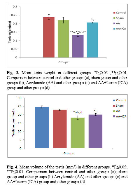
The number of sertoli, spermatogonia, spermatocyte, spermatid, and leydig cells was estimated using the physical dissector counting techniques. A dissector is a 3D sampling probe consists of two parallel planes for counting the number of cells. The size, shape, or spatial position of the cells do not affect this method. Two consecutive sections are then compared. These consecutive sections were chosen randomly. The first chosen section called the reference section and its next section called lookup section, and also called a dissector pair, 30μ separates these used sections as physical dissector pairs. A rule for the dissector counting method is that the distance between two section planes pairs must be about 1/3 of the average interest particle under investigation (in this study cell nuclei) to be estimated. During this method, twenty consecutive dissector pairs from any biopsy were used to the investigation [22]. Only if the cells were not present in the lookup section, and cells do not touch forbidden lines, they would be counted (Fig. 2).
  
 
 To estimate the mean numerical density of cells, the following formula was used:
NV= ΣQ/t × a (p) × h × p
ΣQ; the total number of the counted cells in the reference section, t; the mean section thickness (5μ), a (p); the area of the unbiased counting frame, h; the distance between sections, P; the number of frame associated points hitting the tissue.
To estimate the total number of cells in a whole mice testis, the following equation was used:
Ntotal = NV × Vtotal
NV is the numerical density of cells, Ntotal; the total number of cells in the whole testis calculated using the testis volume results estimated by the Cavalieri method [22].
Assay histopathology changes of prostate
The 5 µm thickness sections were extracted from each prostate and stained with hematoxylin and eosin. The stained sections were examined with the light microscope (Olympus Japan) with the magnification of 100× and 400×. The average area of each of the alveoli and the height of epithelial cells were subsequently measured.
Statistical analysis
Data were statistically analyzed by one-way analysis of variance (ANOVA) followed by Tukey's test. The value of p≤ 0.05 was used as the criterion for statistical significance. All data were expressed as mean ± standard error.

Results
The mean testis weight (mg) in different groups
Significant reductions were seen in the testis weight of Acrylamide treated group compared to control and sham groups (p≤0.01). The testis weight of the Acrylamide+Icariin group was significantly higher than Acrylamide treated group (p≤0.05). There were no significant differences in Acrylamide+Icariin group compared with the control and sham group (Fig. 3).
The mean total volume of testis in different groups
The total volume of testis showed a significant reduction in Acrylamide group compared to the sham and control group (P≤0.05). This parameter in the Acrylamide+Icariin group has increased in comparison with Acrylamide group (P≤0.05). There was no significant difference in Acrylamide+Icariin group compared with the sham and control group (Fig. 4).
Estimated the number of spermatogonia, spermatocyte, spermatid, and sertoli and leydig cells
The total number of spermatogonia cells in Acrylamide group showed a significant decrease in comparison with control, sham, and Acrylamide+Icariin groups (P≤0.05). There was no significant difference in Acrylamide+Icariin group compared with the control and sham group.
The total number of spermatocyte cells in Acrylamide group showed a significant decrease in comparison with control, sham, and Acrylamide+Icariin groups (P≤0.05). There was no significant difference in Acrylamide+Icariin group compared with the control and sham group. The total number of spermatid cells in Acrylamide group showed a significant reduction in comparison with the control and sham group (P≤0.05). The number of this cell was increased in the Acrylamide+Icariin group in comparison with Acrylamide treated group, but it was not statistically significant. There was no significant difference in Acrylamide+Icariin group compared with the control and sham group.
The total number of sertoli cells in Acrylamide group showed a reduction in comparison with the control and sham group, but it was not statistically significant. The number of this cell was increased in the Acrylamide+Icariin group in comparison with Acrylamide treated group, but it was not statistically significant. There was no significant difference in Acrylamide+Icariin group compared with the control and sham group. The number of testicular leydig cells in Acrylamide group showed a significant decrease in comparison with control, sham, and Acrylamide+Icariin groups (P≤0.05). There was no significant difference in Acrylamide+Icariin group compared with the control and sham group (Fig. 5. and Table1).
Johnsen score results
In the histopathologic evaluation, the mean Johnsen score was decreased in the Acrylamide treated group compared to control, sham, and Acrylamide+Icariin groups (P≤0.05). There was no statistically significant difference in the Acrylamide+Icariin group in comparison with the control and sham group (Fig. 6).
 
  
Table 1. Estimated number of spermatogonia, spermatocyte, spermatid, and Sertoli and Leydig cells in different groups
  Groups
Parameters (N) Control Sham Acrylamide Acrylamide + Icariin
Spermatogonia 6.21±0.63 6.95±0.30 4.00±0.75*a,b,d 6.14±0.41*c
Spermatocyte 16.300±1.78 15.45±1.38 9.39±0.92*a,b,d 15.89±1.76*c
Spermatid 8.00±1.46 6.51±0.68 2.75±0.64*a,b 5.25±0.94
Sertoli 1.22±0.160 1.32±0.159 0.83±0.120 1.162±0.07
Lydig 3.19±0.41 3.08±0.55 1.29±0.18*a,b,d 3.02±0.37*c
The results are shown as mean±SEM. P≤0.05 is indicated by"*". Comparison between control and other groups (a), sham group and other groups (b) Acrylamide and other groups (c)and Acrylamide+Icariin group and other groups (d)
 
Testosterone concentration
Testosterone concentration in the Acrylamide group showed a significant decrease in comparison with control, sham, and Acrylamide+Icariin groups (p≤0.05). There was no significant difference in the Acrylamide+Icariin group compared with the control and sham group (Fig. 7).
Histological findings in the testis
In the present study, normal histopathologic features were observed in the control group. There was a normal height of the germinal epithelium. The examined specimens of the testes of the control group showed the seminiferous tubules that were separated by intervening connective tissue containing leydig cells, blood vessels, and other components of connective tissue. Close examination of the wall of the seminiferous tubules showed that it consisted of germinal epithelium supported by Sertoli cells. The germinal epithelium comprised different stages of the spermatogenic series, namely spermatogonia, spermatocytes, and spermatids arranged (Fig 5A).
No obvious histopathological changes were observed in the sham group. The results showed that the seminiferous tubules of the sham group had regular size and cytoarchitecture, and all cells of the spermatogenic series were presented (Fig 5B).
Different degrees of destruction of the seminiferous tubules were demonstrated in the Acrylamide treated group. The results showed that the seminiferous tubules of the Acrylamide treated group had an irregular size, distorted shape, and abnormal cytoarchitecture. In the testes of Acrylamide treated group, some detached and pyknotic round spermatids or retained elongated spermatids were counted. The testes in this group had a significant reduction in spermatids. Also, there was a decrease in height of the germinal epithelium in some slides of this group (Fig. 5C).
The Acrylamide+Icariin group had a normal height of germinal epithelium. The germinal epithelium comprised different stages of the spermatogenic series and sertoli cells. In this group, seminiferous tubules were separated by intervening connective tissue containing Leydig cells (Fig. 5D). The interstitial space between the seminiferous tubules was increased, and were observed in some slides and displacement of sertoli and germinal cells (Figs. 8, 9, 10).
No histopathological changes were seen in the prostatic glands of the control group. Epithelial and stromal had normal structures in the control group (Fig. 11A). The microscopic sections of prostate variations, including a degree of dilatation, was showed in the prostatic gland as well as of their intraluminal secretions. Normal histopathologic features were observed in the sham group (Fig. 11B). All of the prostates from the Acrylamide treated group presented various degrees of epithelial and stromal hyperplasia and epithelial cell proliferation in the prostatic lobes (Fig. 11C). In Acrylamide+Icariin groups, the epithelium of glands has normal shape and size. Epithelial and stromal had a normal structure in this group (Fig. 11D). Epithelium height in the Acrylamide group showed a decrease in comparison with control, sham, and Acrylamide+Icariin groups, but there was no significant difference in different groups (Fig. 8).
 
 A                                              B                                          C                                                D


Discussion
The present study showed that Acrylamide decreased the testis weight and total volume of testis compared to control and sham groups. The total number of spermatogonia cells and spermatocyte cells in the Acrylamide group showed a significant decrease. The total number of spermatid cells in the Acrylamide group showed a significant reduction in comparison with the Acrylamide+Icariin group. The total number of Sertoli cells in the Acrylamide group showed a reduction in comparison with control, sham, and Acrylamide+Icariin groups, but it was not statistically significant. The number of leydig cells in the Acrylamide group showed a significant decrease in comparison with Acrylamide+Icariin groups. Also, the mean Johnsen score was decreased in the Acrylamide treated group compared to Acrylamide+Icariin groups. Besides, testosterone concentration in the Acrylamide group had a reduction in comparison with Acrylamide+Icariin groups.
The decrease in the testis weight and testis volume of the Acrylamide treated group observed in this study agrees with earlier reports. Hye yang et al. [6] have examined six groups of male rats that received Acrylamide at doses of 0, 5, 15, 30, 45, or 60 mg/kg/ day for 5 consecutive days. They observed significant dose-related reduction in testis weight of male rats.
Previous animal studies also demonstrated that Acrylamide reduced the testis volume and weight. Besides, decreased in testis weight and volume of Acrylamide treated group might be due to a decreased number of germ cells because the weight and volume of the testis are mainly dependent on the mass of the differentiated spermatogenic cells [23]. Also, some studies demonstrated that Acrylamide exposure inhibited differentiation of the seminiferous tubules [24], decreased the diameter of seminiferous tubules [25], and the decrease in these cases lead to reductions in testis weight and volume.
In the current study, it is revealed that Acrylamide decreased the number of spermatogonia, spermatocyte, spermatid, sperm, and Sertoli cells. Several animal studies have also demonstrated a negative influence of Acrylamide on the number of spermatogonia and Sertoli cells [6, 26]. In fact, Acrylamide  can reduce the performance of spermatogenesis in male rats by affecting the degeneration of Sertoli cells [25].
In the present study, the histopathologic evaluation showed that the mean Johnsen score was decreased in the Acrylamide treated group compared to the control group. Some studies demonstrated that Acrylamide consumption causes arrest in spermatogenesis and indicated that Johnsen scores decrease due to Acrylamide administration [24, 25]. Zaki et al. [27] studied the protective effect of tomato and carrot against Acrylamide on histopathological sections of some organs in mice. The prostate gland of mice from the Acrylamide treated group revealed atrophy of epithelial lining, necrosis, and fibrosis of epithelium, interstitial edema, and desquamated epithelium in the lumen. In the current study, the effects of Acrylamide consumption on the prostate gland of males were examined. The results showed various degrees of epithelial, stromal hyperplasia, and epithelial cell proliferation in the prostatic lobes in the Acrylamide treated group. Also, Epithelium height in the Acrylamide group showed a decrease in comparison with other groups, which is in line with Zaki's study.
Abdelghaffar et al. [28] have examined three groups of male rats that two of them were received Acrylamide for 28 consecutive days. They observed a significant reduction in testosterone concentration of male rats. In the present study, it is observed that testosterone concentration in the Acrylamide group showed a significant decrease in comparison with control, sham, and Acrylamide+Icariin groups.
The present study showed that Acrylamide+ Icariin group increased testis weight significantly. The total number of spermatogonia cells and spermatocyte cells was increased in the Acrylamide+Icariin group. The total number of spermatid cells was increased in the Acrylamide+Icariin group in comparison with the Acrylamide treated group, but it was not significant. In the present study, it is observed that Icariin consumption causes an increase in testicular weight and volume. Ding et al. [29] reported that Icariin treatment causes a significant dose-dependent increase in the testicular indices in all three Icariin groups.
Icariin administration alongside Acrylamide significantly increased the decreased seminiferous tubule diameters and total Johnson's due to Acrylamide administration. Also, the increase in the number of spermatogenic cells of Acrylamide+Icariin treated group that observed in this study aligns with the earlier reports. Some of the studies demonstrated that Icariin has protective effects on the reproductive system by reduced apoptosis in spermatogenic cells [30, 31].
On the other hand, Xu et al. [32] have examined three groups of male rats that received Icariin at different doses. They reported that exposure to Icariin caused a significant increase in Johnsen's score of the testis. Also, in the present study, it is observed that Icariin consumption causes an increase in Johnsen's score. In Acrylamide+Icariin group epithelium of glands has normal shape and size. Moreover, epithelial and stromal had normal structure in this group.

Conclusion
In conclusion, exposure of mice to Acrylamide may impact gonadal function, and it is confirmed that Acrylamide caused testicular oxidative stress with destructive histopathological changes and reduction in testosterone concentration. Our results demonstrated that Acrylamide exposure had irreversible effects on the testis performance of male rats and altered the testicular microarchitecture and also slowed the negative effects on germ cell proliferation. From the present results, it could be concluded that Icariin can compensate for the toxic effect of Acrylamide on histomorphometric changes of testis and prostate by its antioxidant effects.
Conflict of Interest
The authors declared no conflic of interest.
Acknowledgments
This paper is issued from the MSc thesis of Mahsa Doctor Arastouye Marandi and financially supported by Shahid Sadoughi university of medical sciences, Yazd, Iran. The authors would like to thank the Department of Anatomical Sciences and the faculty of medicine for their official cooperation.


References
  1.  Mustafa HN. Effect of acrylamide on testis of albino rats. Ultrastructure and DNA cytometry study. Saudi Med J. 2012; 33(7): 722-31.
  2.  Tyl RW, Marr MC, Myers CB, Ross WP, Friedman MA. Relationship between acrylamide reproductive and neurotoxicity in male rats. Reproductive Toxicology 2000; 14(2): 147-57.
  3.  Kermani-Alghoraishi M, Anvari M, Talebi AR, Amini-Rad O, Ghahramani R, Miresmaili SM. The effects of acrylamide on sperm parameters and membrane integrity of epididymal spermatozoa in mice. Europ J Obstetric Gynecol Reproduct Biol. 2010; 153(1): 52-5.
  4.  Yang HJ, Lee SH, Jin Y, Choi JH, Han DU, Chae C, et al. Toxicological effects of acrylamide on rat testicular gene expression profile. Reproduct Toxicol. 2005; 19(4): 527-34.
  5.  Wang H, Huang P, Lie T, Li J, Hutz RJ, Li K, et al. Reproductive toxicity of acrylamide-treated male rats. Reproduct Toxicol. 2010; 29(2): 225-30.
  6. Yang HJ, Lee SH, Jin Y, Choi JH, Han CH, Lee MH. Genotoxicity and toxicological effects of acrylamide on reproductive system in male rats. Journal of veterinary science. 2005; 6(2): 103-109.
  7. Weiss G. Acrylamide in food: Uncharted territory. American Association for the Advancement of Science 2002(2): 27.
  8.  Richmond P, Borrow R. Acrylamide in food. Lancet 2003; 361(9355): 361-62.
  9. Hsieh TP, Sheu SY, Sun JS, Chen MH, Liu MH. Icariin isolated from Epimedium pubescens regulates osteoblasts anabolism through BMP-2, SMAD4, and Cbfa1 expression. Phytomedicine 2010; 17(6): 414-23.
  10.  Fan JJ, Cao LG, Wu T, Wang DX, Jin D, Jiang S, et al. The dose-effect of icariin on the proliferation and osteogenic differentiation of human bone mesenchymal stem cells. Molecules 2011; 16(12): 10123-133.
  11. Low WY, Tan HM. Asian traditional medicine for erectile dysfunction. Journal of Men's Health and Gender 2007; 4(3): 245-50.
  12.  Liu T, Xin H, Li WR, Zhou F, Li GY, Gong YQ, et al. Effects of icariin on improving erectile function in streptozotocin‐induced diabetic rats. The Journal of Sexual Medicine. 2011; 8(10): 2761-772.
  13.  Chen M, Hao J, Yang Q, Li G. Effects of icariin on reproductive functions in male rats. Molecules. 2014; 19(7): 9502-514.
  14.  Pourentezari M, Talebi A, Abbasi A, Khalili MA, Mangoli E, Anvari M. Effects of acrylamide on sperm parameters, chromatin quality, and the level of blood testosterone in mice. Iranian Journal of Reproductive Medicine 2014; 12(5): 335.
  15.  Hopwood, D. Fixation and fixatives in theory and practice of histological techniques, Bancroft J, Stevens A ed. London: Churchill Livingstone; 1996.
  16. Guneli E, Tugyan K, Ozturk H, Gumustekin M, Cilaker S, Uysal N. Effect of melatonin on testicular damage in streptozotocin-induced diabetes rats. Europ Surgic Res. 2008; 40(4): 354-60.
  17.  Yadegari M, Khazaei M, Anvari M, Eskandari M. Prenatal caffeine exposure impairs pregnancy in rats. International J Fertil Steril. 2016; 9(4): 558.
  18.  Johnsen SG. Testicular biopsy score count–a method for registration of spermatogenesis in human testes: normal values and results in 335 hypogonadal males. Hormone Research in Paediatrics 1970; 1(1): 2-25.
  19.  Dehghani F, Hassanpour A, Poost-pasand A, Noorafshan A, Karbalay-Doust S. Protective effects of L-carnitine and homogenized testis tissue on the testis and sperm parameters of busulfan-induced infertile male rats. Iran J Reproduct Med. 2013; 11(9): 693.
  20.  Mahmoudi R, Honarmand Z, Karbalay-Doust S, Jafari-Barmak M, Nikseresht M, Noorafshan A. Using curcumin to prevent structural impairments of testicles in rats induced by sodium metabisulfite. EXCLI J. 2017; 16(1): 583.
  21.  Akosman MS, Lenger Ö, Demirel H, Akosman M, Lenger O, Demirel H. Morphological, stereological and histometrical assessment of the testicular parameters between holstein and simmental bulls. Int J Morphol. 2013; 31(3): 1076-80.
  22. Bertram JF. Analyzing renal glomeruli with the new stereology. International review of cytology 1995; 161(1): 111-72.
  23.  Mruk DD, Cheng CY. Sertoli-Sertoli and Sertoli-germ cell interactions and their significance in germ cell movement in the seminiferous epithelium during spermatogenesis. Endocrine Rev. 2004; 25(5): 747-806.
  24. Hasanin NA, Sayed NM, Ghoneim FM, Al-Sherief SA. Histological and ultrastructure study of the testes of acrylamide exposed adult male albino rat and evaluation of the possible protective effect of vitamin E intake. Journal of Microscopy and Ultrastructure 2018; 6(1): 23.
  25.  Erdemli Z, Erdemli ME, Turkoz Y, Gul M, Yigitcan B, Gozukara Bag H. The effects of acrylamide and Vitamin E administration during pregnancy on adult rats testis. Andrologia 2019; 51(7): 13292.
  26.   Sakamoto J, Kurosaka Y, Hashimoto K. Histological changes of acrylamide-induced testicular lesions in mice. Exper Moleculr Pathol. 1988; 48(3): 324-34.
  27.  Zaki SA, Amin WS, Nagi HM. The functional role of tomato and carrot on histopathological lesions of brain, small intestine and prostate in mice treated with acrylamide. Integr Food Nutr Metab. 2014; 1(1): 114-18.
  28. Abdelghaffar S, Omar H, Fiedan I, Ahmed E. Acrylamide induced testicular toxicity in rats: Protective effect of garlic oil. Reproduction in Domestic Animals 2015; 50(1): 1-10.
  29.  Ding J, Tang Y, Tang Z, Zu X, Qi L, Zhang X, et al. Icariin improves the sexual function of male mice through the PI 3K/AKT/eNOS/NO signalling pathway. Andrologia 2018; 50(1): 12802.
  30.  Liu ZP, Li ZX, Pan XY. Effect of icariin on apoptosis of spermatogenic cells in alcohol-treated testis of mice. Maternal and Child Health Care of China 2014; 15(1): 56.
  31. Yuan D, Wang H, He H, Jia L, He Y, Wang T, et al. Protective effects of total flavonoids from Epimedium on the male mouse reproductive system against cyclophosphamide‐induced oxidative injury by up‐regulating the expressions of SOD3 and GPX1. Phytotherapy Research 2014; 28(1): 88-97.
  32.  Xu Yp, Liu Bx, Zhang Xp, Yang Cw, Wang Ch. A Chinese herbal formula, Wuzi Yanzong Pill, improves spermatogenesis by modulating the secretory function of Sertoli cells. Chinese Journal Of Integrative Medicine 2014; 20(3): 194-99.

 
Type of Study: Research | Subject: Pathology
Received: 2020/01/25 | Accepted: 2020/05/15 | Published: 2020/08/27

Add your comments about this article : Your username or Email:
CAPTCHA

Send email to the article author


Rights and permissions
Creative Commons License This work is licensed under a Creative Commons Attribution-NonCommercial 4.0 International License.

© 2024 CC BY-NC 4.0 | International Journal of Medical Laboratory

Designed & Developed by : Yektaweb