Introduction
The kidney can reabsorb and collect divalent metals, so the first target organ is the toxicity of heavy metals [
1,
2]. Mercury is one of the toxic metals that has attracted special attention; the forms of mercury are highly nephrotoxic [
3]. Mercury increases reactive oxygen species such as superoxide anion, hydrogen peroxide, and hydroxyl radicals, which stimulate lipids, proteins, and DNA oxidation, resulting in cell death [4]. Concomitant use of various herbs with anti-oxidant and nephroprotective effects can reduce the toxicity of heavy metals [
5]. Anti-oxidants are one of the essential components in the treatment of mercury poisoning [
4]. Mercuric chloride increases the production of oxidants such as hydrogen peroxide, which can be reduced by protective anti-oxidants such as glutathione (GPX), superoxide dismutase (SOD), or even N-acetyl cysteine [
6]. Evaluation of renal toxicity can be detected through a simple blood test that includes measurement of blood urea nitrogen (BUN), serum creatinine concentration, and glomerular filtration rate [
7]. Serum creatinine is a measure of kidney damage in clinical trials [
8]. An imbalance between free radicals and antioxidant enzymes is called oxidative stress. Reactive oxygen species (ROS) or free radicals can be produced by the cell's natural metabolisms and react with biological molecules such as proteins, lipids, and DNA, causing cell damage and changes in DNA [
9-11]. Oxidative stress plays an important role in developing chronic and degenerative diseases such as cancer, arthritis, aging, autoimmune disorders, cardiovascular, neurological, and renal diseases. ROS and reactive nitrogen species (RNS) are produced from endogenous or exogenous sources. Endogenous free radicals from activated immune cells, inflammation, mental stress, excessive exercise Ischemia, infection, cancer, aging Exogenous ROS / RNS due to air and water pollution, cigarette smoke, alcohol, heavy metals (Cd, Hg, Pb, Fe, As), certain drugs (cyclosporine, tacrolimus, Gentamicin, bleomycin), industrial solvents, cooking meat, fat and radiation [
12,
13].
Any substance that can eliminate active oxygen species or inhibit their production is called an anti-oxidant [
9,
14,
15]. The human body copes with anti-oxidants that are naturally produced or enter the body by foods or supplements through various mechanisms to deal with oxidative stress and reduce the risk of disease [
9,
12]. Classification of anti-oxidants
I. Based on the location: Plasma anti-oxidants: uric acid, bilirubin, transferrin, ceruloplasmin. Cell membrane anti-oxidants: α-Tocopherol. Intracellular anti-oxidants: SOD, catalase (CAT) and GPX, and glutathione reductase.
II. By nature and practice: enzymatic anti-oxidants: SOD, CAT and GPX, and glutathione reductase.
Non-enzymatic antioxidants and nutrient antioxidants: beta-carotene, α-tocopherol, ascorbic acid (Vit C), and vitamin E metabolic anti-oxidants: GSH, bilirubin, uric acid, transferrin, ceruloplasmin, albumin [
11,
16]. Thyme contains large amounts of monoterpenes (thymol, carvacrol, geraniol, α-terpinol, sabin hydrate, linalool, 1 and 8-cineole) terpenoids, flavonoids (quercetin, luteolin, apigenin), glycosides, and phenolic acid (rosmarinic acid) [
17-19]. Reports indicate that the medicinal effects of thyme are due to compounds such as thymol and carvacrol [
20]. Thymol and carvacrol have a synergistic effect on kidney protection, although the protective effect of thymol is better than carvacrol [
21,
22]. Applications of thymol include anti-oxidants, anti-inflammatory, antibacterial, antiviral, antifungal, anti-epileptic, and seizure, reduce respiratory problems, wound healing, improve digestion, reduce menstrual pain [
23-26]. Thymol is also used to treat inflammatory diseases, immune disorders related to oxidative stress [
27]. Also, its anti-oxidant effect can increase the activity of antioxidants such as SOD and GPX and delay lipid oxidation [
18,
22,
28].
Materials and Methods
Thymol, mercuric chloride, Catalase Assay kit, SOD, and GPX assay kit were purchased from Sigma Aldrich Company. Assay for urea and creatinine were conducted with a standard test at the laboratory of Yazd Diabetic center. In this study, about 30 male rats (weighing approximately 250-300 g) were purchased from the infertility center in Yazd and kept at a temperature of 22
C ° with sufficient light (12 hours of light and 12 hours of darkness). In order to adapt to the new environment of rats, they were kept for ten days. They were kept in the pet house of the Faculty of Shahid Sadoughi University of Medical Sciences, Yazd, in large cages made of sterile polyethylene glycol. Thirty rats were divided into six groups of five: the first group, as the non-exposed group (Intact), who received corn oil as thymol solvent for 15 days. The second group, or control group, animals that received mercury chloride at a single dose of 0.5 mg/kg for 15 days. The third group was animals that received mercury chloride at a dose of 0.5 mg/kg for 15 days and then thymol at a dose of 10 mg/kg intraperitoneally for five days. The fourth group was the animals that received mercury chloride at a dose of 0.5 mg/kg for 15 days and then thymol at 30 mg/kg intraperitoneally for five days. The fifth group, animals that had 15 days of mercury, received thymol at a dose of 50 mg/kg intraperitoneally for five days. Group 6 received mercury chloride at a dose of 0.5 mg/kg for 15 days and then thymol at a dose of 5 30 mg/kg intraperitoneally for ten days. Initially, deep anesthesia was induced by diethyl ether in the animals, and finally, their blood samples were taken from the heart and kidney tissue samples for research and biochemical tests. Blood samples were collected in tubes without anticoagulants and centrifuged at 3000 rpm for 20 minutes to obtain serum. Serum was transferred to clean, dry, labeled microtubes and transferred to a freezer at -20 °C for further biochemical analysis. We also inserted kidney tissue samples into clean and labeled falcons and transferred them to a freezer at -20 °C for further biochemical analysis in the central laboratory of pharmacy school in Yazd. The required amount of serum sample was sent to the laboratory of Yazd Diabetes Center, and the amount of serum urea and creatinine was measured and announced. The manuscript was approved by the Research Deputy and Ethics Committee of Shahid Sadoughi University of Medical Science, Yazd, Iran, before the initiation
of the study (No: IR.SSU.MEDICINE.REC.1398.048).
Statistical analysis
Data were expressed as mean values ± standard error of the mean (SEM). We used one-way ANOVA for the first Figure (parametric data), and for nonparametric data, we used median for data analysis. The value with
P < 0.05 was considered significant.
Results
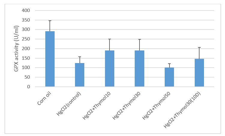
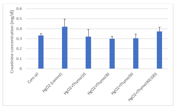
Measurement of catalase activity in kidney tissue sample in Figure 1- Statistically significant corn oil with group received thymol 10 mg/kg dose (p < 0.05) and the activity of this enzyme in the presence of mercury chloride (control) has increased. The activity of superoxide dismutase enzyme in kidney tissue samples in Figure 2, using a median test (p < 0/05). There was a significant difference in superoxide dismutase activity in the corn oil group (intact) and mercury chloride group. The superoxide dismutase activity in the presence of thymol at a dose of 10, 30, and 50 mg/kg was reduced compared to mercury chloride (p < 0.05). The GPX enzyme activity in kidney tissue samples in Figure 3- using the median test, we could not obtain a statistically significant difference due to the small number of samples (p > 0.05). Serum urea concentration in Figure 4- using the median test. There was a significant difference in serum urea concentration in the mercury chloride group, with the group receiving thymol at a dose of 50 mg/kg (#p ≤ 0.05). Serum creatinine concentration in serum sample in Figure 5. Using the median test, we could not obtain a statistically significant difference due to the small number of samples (p > 0.05).
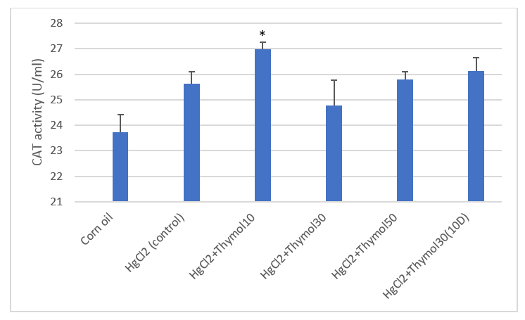
Fig. 1 . Comparison of catalase (CAT) enzyme activity in kidney tissue samples of treated animals. Respectively: groups 1- Corn oil (intact group), 2- mercuric chloride (control group), 3- mercuric chloride + Thymol 10, 4- mercuric chloride + Thymol30 , 5- mercuric chloride + Thymol 50 mg / kg and 6- mercuric chloride + Thymol30 for 10 days. *denotes the result of statistical analysis of the corn oil group, which was statistically significant with the intervention group (*p < 0.05).
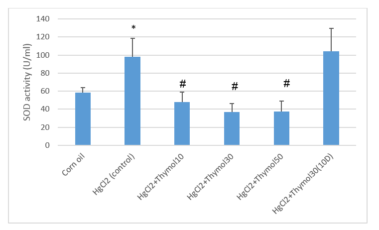
Fig. 2. Comparison of the superoxide dismutase activity (SOD) enzyme activity in kidney tissue samples of treated animals. Respectively: groups 1- Corn oil (intact group), 2- mercuric chloride (control group), 3- mercuric chloride + Thymol 10, 4- mercuric chloride + Thymol 30, 5- mercuric chloride + Thymol 50 mg/kg and 6- mercuric chloride + Thymol30 for ten days. * p ≤ 0.05, # denotes the result of statistical analysis of the control group, which was statistically significant with the intervention groups (#p ≤ 0.05).
Fig. 3. Comparison of glutathione peroxidase (GPX) enzyme activity in kidney tissue samples of treated animals in intervention and (mg/kg) control groups. Respectively: groups 1- Corn oil (intact group), 2- mercuric chloride (control group), 3- mercuric chloride + Thymol 10, 4- mercuric chloride + Thymol 30, 5- mercuric chloride + Thymol 50 mg/kg and 6- mercuric chloride + Thymol 30 for 10 days.
Fig. 4. Comparison of the urea concentration in serum samples of treated animals, respectively: groups 1- Corn oil (intact group), 2- mercuric chloride (control group), 3- mercuric chloride + Thymol 10, 4- mercuric chloride + Thymol 30, 5- mercuric chloride + Thymol 50 mg / kg and 6- mercuric chloride + Thymol30 for 10 days. # denotes intervention groups with the control group, was statistically significant (#p

0.05)
.
Fig. 5. Comparison of the creatinine concentration in serum samples of treated animals in intervention and (mg/kg) control groups. Respectively: groups 1- Corn oil (intact group), 2- mercuric chloride (control group), 3- mercuric chloride + Thymol 10, 4- mercuric chloride + Thymol30, 5- mercuric chloride + Thymol 50 mg/kg and 6- mercuric chloride + Thymol30 for 10 days. There is no significance within and between groups (p > 0.05).
Discussion
There is a significant difference in the activity of catalase enzyme in kidney tissue samples in the group of corn oil (intact) with the group that received thymol at a dose of 10 mg/kg, and the highest activity of catalase enzyme is visible in this group, so thymol with the maximum role of anti-oxidant has been able to counteract the oxidants of mercury chloride. In general, at low concentrations of thymol, the activity of catalase in the cell increases and helps the cell fight the oxidative state of mercury chloride. It may be due to a change in gene expression or the OH functional group, which acts like vitamin E. Vitamin E is an anti-oxidant in low concentrations. The activity of the superoxide dismutase enzyme in the group of mercury chloride has increased compared to the group of corn oil, which is similar to the activity of the enzyme catalase, because when we give mercury chloride, the activity of free radicals increases, so the enzyme superoxide dismutase activity was increasing to reduce the effect of oxidants, as has been shown in other studies [
29]. Also, by giving doses of 10, 30, and 50 mg/kg thymol, the superoxide dismutase enzyme activity decreased compared to the control group because thymol reduced the free radicals caused by mercury chloride, so the activity of the superoxide dismutase enzyme also decreased. In other studies, this result was obtained [
29,
30]. As the thymol concentration increases, it has a negative effect on the activity of the superoxide dismutase enzyme. In general, it can be concluded that the mechanism of action of catalase is different from that of superoxide dismutase, even though we do not know the exact mechanism of these enzymes. As mentioned in chapter 2, the enzyme catalase inactivates oxygenated water

, and the enzyme superoxide dismutase inactivates the superoxide anion (O
−2) (11). As shown in the diagrams of catalase and superoxide dismutase results, the activity of superoxide dismutase decreased in the groups that received thymol, so it can be said that thymol, as an anti-oxidant, was able to reduce the superoxide anion (O
−2). As a result, mercury chloride was able to produce more superoxide anion (O
−2) so thymol, as an anti-oxidant, was also able to cause more reduction of superoxide anion (O
− 2), so the activity of the superoxide dismutase enzyme is reduced in these groups. As can be seen in the results of GPX activity, there are clinically significant differences between the groups, and they seem significant, but statistically, due to the small number of samples, we could not achieve a significant level [
29,
31,
32]. Also, by giving different doses of thymol, the activity of GPX has increased compared to the control group because thymol has anti-oxidant activity and can increase the activity of GPX [
31,
33]. However, the group receiving thymol at a dose of 50 mg/kg not only did not increase the activity of GPX but also decreased the activity of this enzyme. Therefore, low thymol doses generally increase GPX activity, but they are not significant, as can be seen from the urea concentration results. Clinically, we expected the urea concentration to be higher in the group receiving mercury chloride (control) than in the corn oil (intact) group, but there was not much difference that could be concluded that urea was not significant. In this study, the urea concentration in the group receiving thymol at a dose of 50 mg/kg was significantly different from the control group because thymol could reduce the renal toxicity of mercury chloride by increasing its antioxidant activity [
29].
As can be seen in the results of creatinine concentration, we expect creatinine concentration to increase in the mercury chloride group (control), which has an oxidizing role compared to the corn oil group (control) because mercury chloride plays an oxidant role in renal toxicity. Serum creatinine increases with damage to the kidneys [
31]. However, in terms of statistical data in this study, no significant difference was observed due to the small number of samples. Considering that the creatinine concentration in the mercury chloride group has increased, that can be because of kidney damage. Creatinine is an excretory substance, and it enters the urine through diffusion; it can no longer return to the blood unless it can return to the blood through kidney damaged. Also, by giving different doses of thymol, the creatinine concentration decreased compared to the control group. By increasing its anti-oxidant activity, thymol tries to reduce the effect of renal toxicity caused by mercury chloride and thus reduce serum creatinine [
31]. The highest concentration of creatinine belongs to the control group, i.e., mercury chloride as an oxidant could have a toxic effect on the kidneys, so serum creatinine is increased, but there is no significant difference between the control and treated groups.
Conclusion
Based on the evidence from this study, it can be concluded that mercury chloride causes oxidative stress and thus increases the production of oxidants and damage to kidney tissue. Thymol in different doses can reduce the renal toxicity caused by mercury chloride. Due to its anti-oxidant effect, thymol increased the activity of antioxidants such as catalase and delayed oxidative stress, and the destruction of kidney tissue. The GPX activity seems to be significant due to the small number of samples. No significant difference showed in the data. SOD had different change than other enzymes because it significantly decreased in animal treated with different doses of thymol that can be because of the protective effect of thymol on this enzyme so we can say the reduction of SOD protect the kidney from damage induced by mercury chloride. On the other hand, 50 mg/kg of thymol significantly reduced urea concentration because of its protective effect on the kidney.
Conflicts of Interest
The authors declare no conflict of interest.
Acknowledgment
None.
References
- Barbier O, Jacquillet G, Tauc M, Cougnon M, Poujeol P. Effect of heavy metals on, and handling by, the kidney. Nephron Physiol. 2005; 99(4): 105-110.
- Agha FE, Youness ER, Selim MM, Ahmed HH. Nephroprotective potential of selenium and taurine against mercuric chloride induced nephropathy in rats. Renal Fail. 2014; 36(5): 704-16.
- Bridges CC, Zalups RK. The aging kidney and the nephrotoxic effects of mercury. J Toxic and Environ Hlth. 2017; 20(2): 55-80.
- Augusti PR, Conterato GM, Somacal S, Sobieski R, Spohr PR, Torres JV, et al. Effect of astaxanthin on kidney function impairment and oxidative stress induced by mercuric chloride in rats. Food Chem Toxicol. 2008; 46(1): 212-16.
- Gaikwad K, Dagle P, Choughule P, Joshi YM, Kadam V. A review on some nephroprotective medicinal plants. Int J Phamaceut Sci Res. 2012; 3(8): 2451.
- El-Shenawy SM, Hassan NS. Comparative evaluation of the protective effect of selenium and garlic against liver and kidney damage induced by mercury chloride in the rats. Pharmacological reports. 2008; 60(2): 199.
- Kim SY, Moon A. Drug-induced nephrotoxicity and its biomarkers. Biomol Ther. 20 (3): 268-72.
- Ferguson MA, Vaidya VS, Bonventre JV. Biomarkers of nephrotoxic acute kidney injury. Toxicology 2008; 245(3): 182-93.
- Erejuwa OO, Sulaiman SA, Wahab MS. Honey: a novel antioxidant. Molecules 2012; 17(4): 4400-423.
- Pham-Huy LA, He H, Pham-Huy C. Free radicals, antioxidants in disease and health. Int J biomed Sci. 2008; 4(2): 89-98.
- Mut-Salud N, Álvarez PJ, Garrido JM, Carrasco E, Aránega A, Rodríguez-Serrano F. Antioxidant intake and antitumor therapy: toward nutritional recommendations for optimal results. Oxid Med Cell Longevity. 2016; 2016(1): 6719534.
- Aboonabi A, Rahmat A, Othman F. Antioxidant effect of pomegranate against streptozotocin-nicotinamide generated oxidative stress induced diabetic rats. Toxicol Rep. 2014; 1(9): 915-22.
- Valko M, Leibfritz D, Moncol J, Cronin MT, Mazur M, Telser J. Free radicals and anti-oxidants in normal physiological functions and human disease. Int J Biochem Cell Biol. 2007; 39(1): 44-84.
- Rašković A, Pavlović N, Kvrgić M, Sudji J, Mitić G, Čapo I, Mikov M. Effects of pharmaceutical formulations containing thyme on carbon tetrachloride-induced liver injury in rats. BMC Compl Alternat Med. 2015;15(1):1-10.
- Nagoor Meeran MF, Javed H, Al Taee H, Azimullah S, Ojha SK. Pharmacological properties and molecular mechanisms of thymol: prospects for its therapeutic potential and pharmaceutical development. Front Pharmacol. 2017; 8(3): 380-89.
- Bacova K, Zitterl-Eglseer K, Chrastinova L, Laukova A, Madarova M, Gancarcikova S, et al. Effect of thymol addition and withdrawal on some blood parameters, antioxidative defence system and fatty acid profile in rabbit muscle. Animals 2020; 10(8): 1248.
- Gumus R, Ercan N, Imik H. The Effect of Thyme Essential Oil (Thymus Vulgaris) Added to Quail Diets on Performance, Some Blood Parameters, and the Antioxidative Metabolism of the Serum and Liver Tissues. Revista Brasileira de Ciência Avícola. 2017; 19(2): 297-304.
- El-Sayed EM, Abd-Allah AR, Mansour AM, El-Arabey AA. Thymol and carvacrol prevent cisplatin-induced nephrotoxicity by abrogation of oxidative stress, inflammation, and apoptosis in rats. J Biochem Mol Toxicol. 2015; 29(4): 165-72.
- Youdim KA, Deans SG. Effect of thyme oil and thymol dietary supplementation on the anti-oxidant status and fatty acid composition of the ageing rat brain. Br J Nutrit. 2007; 83(1): 87-93.
- Dhaneshwar S, Patel V, Patil D, Meena G. Studies on synthesis, stability, release and pharmacodynamic profile of a novel diacerein-thymol prodrug. Bioorgan Med Chem Let. 2013; 23(1): 55-61.
- Al-Snafi AE, Thuwaini MM. Nephro- protective effects of Arabian medicinal plants. Res J Pharmaceut, Biologic Chemic Sci. 2018; 9(5): 1504-511.
- Deng LL, Taxipalati M, Que F, Zhang H. Physical characterization and antioxidant activity of thymol solubilized Tween 80 micelles. Scientific Rep. 2016; 6(1): 1-8.
- Chaudhry H, Fatima N, Ahmad IZ. Evaluation of antioxidant and antibacterial potentials of nigella sativa L. suspension cultures under elicitation. Biomed Res Int. 2015; 2015(1): 708691.
- Khazdair MR, Ghorani V, Alavinezhad A, Boskabady MH. Pharmacological effects of Zataria multiflora Boiss L. and its constituents focus on their anti-inflammatory, anti-oxidant, and immunomodulatory effects. Fundament Clinic Pharmacol. 2018; 32(1): 26-50.
- Hashemipour H, Kermanshahi H, Golian A, Veldkamp T. Effect of thymol and carvacrol feed supplementation on performance, anti-oxidant enzyme activities, fatty acid composition, digestive enzyme activities, and immune response in broiler chickens. Poultry Sci. 2013; 92(8): 2059-2069.
- Jamshidi HR, Kalantar H. Effect of sodium selenide on renal toxicity induced by mercuric chloride in rat. Int J MedLab. 2020; 7(2): 90-101.
- Pena C, Hernandez-Fonseca JP, Pedreanez A, Viera N, Mosquera J. Renal oxidative stress and renal CD8(+) T-cell infiltration in mercuric chloride-induced nephropathy in rats: role of angiotensin II. J Immunotoxicol. 2016; 13(3): 324-34.
- Teixeira FB, de Oliveira AC, Leão LK, Fagundes NC, Fernandes RM, Fernandes LM, et al. Exposure to Inorganic mercury causes oxidative stress, cell death, and functional deficits in the motor cortex. Frontiers in molecular neuroscience. 2018; 11(1): 125-35.
- Agarwal R, Behari JR. Role of selenium in mercury intoxication in mice. Industrial Health. 2007; 45(3): 388-95.
- Almeer RS, Albasher G, Kassab RB, Ibrahim SR, Alotibi F, Alarifi S, et al. Ziziphus spina-christi leaf extract attenuates mercury chloride-induced testicular dysfunction in rats. Environm Sci Pollut Res. 2020; 27(3): 3401-412.
- Gao D, Zeng LN, Zhang P, Ma ZJ, Li RS, Zhao YL, et al. Rhubarb anthraquinones protect rats against mercuric chloride (HgCl2)-induced acute renal failure. Molecules 2016; 21(3): 298.
- Gado AM, Aldahm`ash BA. Anti-oxidant effect of Arabic gum against mercuric chloride-induced nephrotoxicity. Drug Design Develop Therap. 2013; 7(1): 245-52.
- Hosseini A, Rajabian A, Fanoudi S, Farzadnia M, Boroushaki MT. Protective effect of Rheum turkestanicum root against mercuric chloride-induced hepatorenal toxicity in rats. Avicenna J Phytomed. 2018; 8(6): 488-98.