
International Journal of Medical Laboratory
Shahid Sadoughi University of Medical Sciences
Wed, Jul 22, 2026
[Archive]

Volume 11, Issue 3 (August 2024)
IJML 2024, 11(3): 202-216 |
Back to browse issues page


Download citation:
BibTeX | RIS | EndNote | Medlars | ProCite | Reference Manager | RefWorks
Send citation to:


BibTeX | RIS | EndNote | Medlars | ProCite | Reference Manager | RefWorks
Send citation to:
Behboudi L, Mahmoudi Hashemi H, Gholami Farashah M S, Pourentezari M, Hashemibeni B, Dortaj H, et al . Utilizing Adipose Derived Stem Cells and Herbal Medicines in Tissue Engineering Approaches
for Cartilage Regeneration. IJML 2024; 11 (3) :202-216
URL: http://ijml.ssu.ac.ir/article-1-522-en.html
URL: http://ijml.ssu.ac.ir/article-1-522-en.html
Leyla Behboudi 
 , Hanie Mahmoudi Hashemi , Mohammad Sadegh Gholami Farashah , Majid Pourentezari * , Batool Hashemibeni , Hengameh Dortaj , Ali Rajabi , Sepideh Izadi
, Hanie Mahmoudi Hashemi , Mohammad Sadegh Gholami Farashah , Majid Pourentezari * , Batool Hashemibeni , Hengameh Dortaj , Ali Rajabi , Sepideh Izadi
Department of Anatomy and Molecular Biology, Shahid Sadoughi University of Medical Sciences, Yazd, Iran & Yazd Neuroendocrin Research Center, Shahid Sadoughi University of Medical Sciences, Yazd, Iran
Full-Text [PDF 336 kb]
(291 Downloads)
| Abstract (HTML) (744 Views)
Coll= Collagen; AGG= Aggrecan; ASU= Avocado soy unsaponifiables; PLGA= Poly lactic-co-glycolic acid; HA= Hyaluronic acid; hADSC= Human adipose derived stem cells; TGF-ß= Transforming growth factor beta; PCR= Polymerase chain reaction; ECM= Extracellular matrix; 3D= Three-dimensional; ICA= Icariin; PFE= Pomegranate fruit extract; CAG= Cycloastragenol; MTT= (3-[4,5-dimethylthiazol-2-yl]-2,5 diphenyl tetrazolium bromide); SOX9= SRY-Box transcription factor 9
Conclusion
References
Full-Text: (288 Views)
Introduction
Tissue engineering is a contemporary discipline focused on developing and regenerating artificial tissues and organs. The concept of replacing damaged body parts with materials derived from natural sources dates back over 4,000 years. However, it has only recently been realized that the engineering of living tissues has led to the establishment of the tissue engineering field. Tissue engineering is an emerging area of research that shows significant potential for replacing damaged tissues by integrating three key components: cells, scaffolds, and bioactive factors [1]. Current research in the field of tissue engineering is centered on advancing the three essential components-cells, scaffolds, and bioactive factors to address fundamental questions and create functional living tissues. Tissue engineering has already achieved significant success in generating avascular tissues and organs, and it holds considerable promise for developing more complex tissues and organs that incorporate highly organized three-dimensional (3D) vascular structures [2-4].
In the future, tissue engineering is anticipated to yield more complex tissues and organs, which could address the shortage of organ donations, decrease the reliance on animal models in drug discovery and toxicity research, and support the advancement of patient-specific smart diagnostics and personalized medicine. In addition to applied tissue engineering, a comprehensive understanding of the fundamental sciences that govern cellular behavior, including their microenvironment and the signaling mechanisms that regulate their functions, is essential [5].
Cartilage Tissue Engineering
Cartilage tissue engineering focuses on creating functional cartilage tissue to replace damaged or lost cartilage. Conventional treatment approaches for cartilage defects frequently fall short of delivering satisfactory results, primarily due to the limited regenerative capacity of cartilage [6]. Tissue engineering strategies address this limitation by employing stem cells and bioactive factors to enhance chondrogenesis. Due to its avascular and aneural characteristics, as well as its composition of a single cell type (chondrocytes), cartilage tissue has emerged as one of the initial candidates for tissue engineering and a prime target for early efforts to create living and functional tissue constructs in vitro. Additionally, the influence of surrounding tissues must be taken into account in orthopedic tissue engineering. While bone and cartilage are distinct tissues, their development is interconnected [7]. The transcription factor Sox9 is expressed in chondrocytes and regulates chondrogenesis. It also suppresses the later stages of osteochondral bone formation by downregulating vasculogenesis [8]. The induction of differentiation in chondrocytes is a critical aspect of cartilage tissue engineering. When cultured in a monolayer, human articular cartilage cells exhibit regular growth and differentiation, simultaneously expressing proteoglycans and type II collagen (Coll II) [9]. Several studies have proposed co-culture systems that combine chondrocytes and stem cells to overcome various challenges associated with monocultures in cartilage tissue engineering. Additional research has indicated that integrating co-culture with 3D biomaterial scaffolds may enhance the effectiveness of these approaches [10]. The advanced strategy of the 3D culture of chondrocytes on hydrogel scaffolds has been shown to prevent chondrocyte dedifferentiation and preserve chondrogenic phenotype [11-13]. 3D scaffolds significantly influence mesenchymal stem cells (MSCs) in their chondrogenesis, as the mechanical environment, hydrostatic pressure, tensile strain, cell-cell interactions, bioactive compound gradients, and other factors created by the cells in 3D culture play a crucial role. These conditions mimic the processes observed during embryonic development, thereby enhancing the differentiation of stem cells [12, 14, 15]. Stem cells possess the ability to differentiate into various tissue-forming cells critical for cartilage and bone regeneration, with MSCs, induced pluripotent stem cells, and embryonic stem cells being the most extensively studied. Due to their superior proliferative capacity compared to chondrocytes, combining stem cells with chondrocyte-derived cells may not accurately represent the optimal cell density required for effective tissue repair. Therefore, it is essential to evaluate the optimal cell density for each specific defect volume while also considering the type of matrix employed. Furthermore, the design and fabrication of scaffolds are integral to tissue engineering, as they establish a three-dimensional microenvironment that facilitates cell attachment, proliferation, differentiation, and the secretion of specific extracellular matrix (ECM) [16].
Adipose Tissue-Derived Stem Cells in Cartilage Tissue Engineering
Adipose tissue-derived stem cells (ADSCs) are multipotent stem cells that can be isolated from adipose tissue. These cells have garnered considerable interest in regenerative medicine due to their abundance, ease of isolation, and capacity to differentiate into various cell types, including chondrocytes [17]. ADSCs have shown great potential for CTE, as they can be readily obtained through minimally invasive procedures such as liposuction [18].
ADSCs can be induced to differentiate into chondrocytes through various methods, including bioactive compounds and herbal medicine [19, 20]. The differentiation process involves expressing specific genes and producing ECM components characteristic of mature cartilage tissue [10]. Moreover, the stromal vascular fraction of adipose tissue has been shown to contain up to 2% of cells capable of differentiating into various cell types, including osteoblasts, chondrocytes, adipocytes, and neurons. In contrast, bone marrow contains only approximately 0.002% of cells with similar differentiation potential [21]. Flow cytometry analysis has been widely used to assess stem cells' surface immunophenotype isolated from humans and other species. Previous studies have demonstrated that human adipose-derived stem cells (hADSCs) express specific adhesion molecules, such as CD9, as well as MSC markers, including CD90, CD44, and CD73, along with the histocompatibility antigen human leukocyte antigens. In contrast, hematopoietic antigens (CD31, CD34, and CD45), the stem cell factor CD117, and the histocompatibility antigen human leukocyte antigens have been identified as absent from the surface of hADSCs [22]. Flow cytometry analysis was conducted using the aforementioned panel of antibodies to evaluate whether hADSCs maintain their stem cell immunophenotype following expansion in culture. Given their ease of collection and ability to undergo multipotential differentiation, hADSCs represent a promising cell source for treating cartilage lesions. These stem cells can be readily expanded in culture over multiple passages to achieve a sufficient and homogeneous population before differentiating into chondrocytes, the cells responsible for cartilage formation [23]. However, the characteristics and differentiation capacity of serially passaged hADSCs have not yet been reported in detail.
Bioactive compounds in cartilage tissue engineering
Bioactive compounds play a vital role in promoting the chondrogenesis of ADSCs. Various anabolic bioactive compounds enhance the synthesis of proteoglycans, aggrecan, and Coll II by chondrocytes, stimulate the proliferation of synoviocytes and MSCs, and promote the chondrogenic differentiation of MSCs. Additionally, catabolic cytokines
such as interleukin-1 (IL)-1 can negatively impact the ECM by increasing matrix metalloproteinase (MMP) activity. These signaling molecules regulate cellular processes and direct stem cells toward a chondrogenic lineage. Commonly utilized growth factors in CTE include transforming growth factor-beta (TGF-β), insulin-like growth factor-1 (IGF-1), and fibroblast growth factor-2 (FGF-2) [24, 25].
TGF-β is recognized for its capacity to induce chondrogenesis and enhance the synthesis of cartilage-specific ECM components. It promotes the expression of chondrogenic markers, including Coll II and aggrecan (AGG), thereby facilitating the formation of functional cartilage tissue. [26, 27]. IGF-1 and FGF-2 also contribute to chondrogenesis by enhancing cell proliferation, matrix synthesis, and cell survival [28].
Growth factors (GFs) are the major regulators of cell behavior. They promote cell proliferation, migration, and differentiation by specific receptor bindings that stimulate cellular signal transduction pathways [29]. GFs involve several physiological and pathological processes, such as tissue repair and hemostasis. GFs can be released from ECM by degrading ECM proteins, GAGs, or PGs [30, 31]. Several GFs and cytokines have been suggested to be involved in chondrogenesis. TGF-βs family includes TGF-β-1, -2, -3, and bone morphogenic proteins (BMPs), which have a prominent role in chondrocyte ECM metabolism and activity as a major inducer of collagen synthesis and tissue homeostasis [19, 32-34]. Most bioactive compounds have been assessed individually rather than in combination to determine their effects on cartilage homeostasis, both in vitro and in vivo. Given the complexity and interactions of these compounds necessary for optimal cartilage growth and maintenance, it is improbable that any single bioactive compound can achieve complete cartilage repair or significantly influence the arthritic environment. Additionally, this protein family uniquely activates SMAD-dependent signaling and transcription and SMAD-independent signaling pathways via MAPKs, such as ERK and TAK1 [35-37]. TGF-β can induce SOX9 synthesis and promote AGG, Coll II, and ECM synthesis by activating the SMAD2/3 phosphorylation pathway, leading to articular cartilage repair [38]. BMPs stimulate cartilage synthesis and decrease the activity of catabolic cytokines, such as IL-1, IL-6, IL-8, MMP-1, and MMP-13 [39]. Moreover, BMP-7 may reduce the degradation of articular cartilage in osteoarthritis [40, 41]. Some studies have investigated new approaches with IGF-I/ FGF-2/ TGF-β/ BMPs/ SOX combinations for cell-based articular cartilage repair [42, 43].
Herbal medicines as bioactive compounds in cartilage tissue engineering
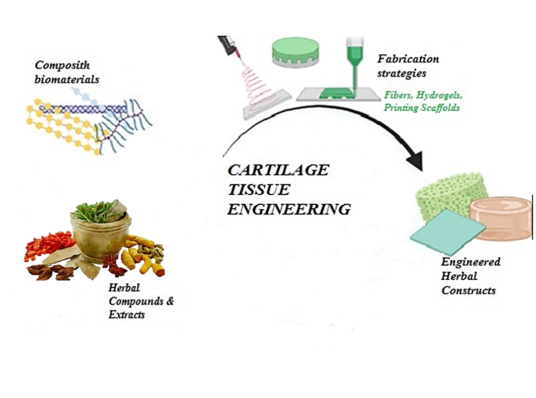
In addition to bioactive compounds, herbal medicine has shown potential in enhancing the chondrogenesis of ADSCs. Extensive screening of traditional medicinal plants has effectively treated infections, diseases, and inflammatory conditions (Fig. 1). Certain herbs possess chondrogenic properties and have been found to stimulate the proliferation of mature stem cells, facilitating the regeneration of damaged tissues. Many Chinese herbs exhibit adipogenic, osteogenic, and chondrogenic effects on human mesenchymal stem cells (hMSCs). Current research is focused on integrating medicinal plant extracts and their bioactive compounds with polymers for tissue regeneration applications. These herbal extracts promote the differentiation of ADSCs into chondrocytes and enhance the production of cartilage-specific extracellular matrix components while reducing inflammation, thereby contributing to effective cartilage tissue engineering strategies (Table 1).
In the future, tissue engineering is anticipated to yield more complex tissues and organs, which could address the shortage of organ donations, decrease the reliance on animal models in drug discovery and toxicity research, and support the advancement of patient-specific smart diagnostics and personalized medicine. In addition to applied tissue engineering, a comprehensive understanding of the fundamental sciences that govern cellular behavior, including their microenvironment and the signaling mechanisms that regulate their functions, is essential [5].
Cartilage Tissue Engineering
Cartilage tissue engineering focuses on creating functional cartilage tissue to replace damaged or lost cartilage. Conventional treatment approaches for cartilage defects frequently fall short of delivering satisfactory results, primarily due to the limited regenerative capacity of cartilage [6]. Tissue engineering strategies address this limitation by employing stem cells and bioactive factors to enhance chondrogenesis. Due to its avascular and aneural characteristics, as well as its composition of a single cell type (chondrocytes), cartilage tissue has emerged as one of the initial candidates for tissue engineering and a prime target for early efforts to create living and functional tissue constructs in vitro. Additionally, the influence of surrounding tissues must be taken into account in orthopedic tissue engineering. While bone and cartilage are distinct tissues, their development is interconnected [7]. The transcription factor Sox9 is expressed in chondrocytes and regulates chondrogenesis. It also suppresses the later stages of osteochondral bone formation by downregulating vasculogenesis [8]. The induction of differentiation in chondrocytes is a critical aspect of cartilage tissue engineering. When cultured in a monolayer, human articular cartilage cells exhibit regular growth and differentiation, simultaneously expressing proteoglycans and type II collagen (Coll II) [9]. Several studies have proposed co-culture systems that combine chondrocytes and stem cells to overcome various challenges associated with monocultures in cartilage tissue engineering. Additional research has indicated that integrating co-culture with 3D biomaterial scaffolds may enhance the effectiveness of these approaches [10]. The advanced strategy of the 3D culture of chondrocytes on hydrogel scaffolds has been shown to prevent chondrocyte dedifferentiation and preserve chondrogenic phenotype [11-13]. 3D scaffolds significantly influence mesenchymal stem cells (MSCs) in their chondrogenesis, as the mechanical environment, hydrostatic pressure, tensile strain, cell-cell interactions, bioactive compound gradients, and other factors created by the cells in 3D culture play a crucial role. These conditions mimic the processes observed during embryonic development, thereby enhancing the differentiation of stem cells [12, 14, 15]. Stem cells possess the ability to differentiate into various tissue-forming cells critical for cartilage and bone regeneration, with MSCs, induced pluripotent stem cells, and embryonic stem cells being the most extensively studied. Due to their superior proliferative capacity compared to chondrocytes, combining stem cells with chondrocyte-derived cells may not accurately represent the optimal cell density required for effective tissue repair. Therefore, it is essential to evaluate the optimal cell density for each specific defect volume while also considering the type of matrix employed. Furthermore, the design and fabrication of scaffolds are integral to tissue engineering, as they establish a three-dimensional microenvironment that facilitates cell attachment, proliferation, differentiation, and the secretion of specific extracellular matrix (ECM) [16].
Adipose Tissue-Derived Stem Cells in Cartilage Tissue Engineering
Adipose tissue-derived stem cells (ADSCs) are multipotent stem cells that can be isolated from adipose tissue. These cells have garnered considerable interest in regenerative medicine due to their abundance, ease of isolation, and capacity to differentiate into various cell types, including chondrocytes [17]. ADSCs have shown great potential for CTE, as they can be readily obtained through minimally invasive procedures such as liposuction [18].
ADSCs can be induced to differentiate into chondrocytes through various methods, including bioactive compounds and herbal medicine [19, 20]. The differentiation process involves expressing specific genes and producing ECM components characteristic of mature cartilage tissue [10]. Moreover, the stromal vascular fraction of adipose tissue has been shown to contain up to 2% of cells capable of differentiating into various cell types, including osteoblasts, chondrocytes, adipocytes, and neurons. In contrast, bone marrow contains only approximately 0.002% of cells with similar differentiation potential [21]. Flow cytometry analysis has been widely used to assess stem cells' surface immunophenotype isolated from humans and other species. Previous studies have demonstrated that human adipose-derived stem cells (hADSCs) express specific adhesion molecules, such as CD9, as well as MSC markers, including CD90, CD44, and CD73, along with the histocompatibility antigen human leukocyte antigens. In contrast, hematopoietic antigens (CD31, CD34, and CD45), the stem cell factor CD117, and the histocompatibility antigen human leukocyte antigens have been identified as absent from the surface of hADSCs [22]. Flow cytometry analysis was conducted using the aforementioned panel of antibodies to evaluate whether hADSCs maintain their stem cell immunophenotype following expansion in culture. Given their ease of collection and ability to undergo multipotential differentiation, hADSCs represent a promising cell source for treating cartilage lesions. These stem cells can be readily expanded in culture over multiple passages to achieve a sufficient and homogeneous population before differentiating into chondrocytes, the cells responsible for cartilage formation [23]. However, the characteristics and differentiation capacity of serially passaged hADSCs have not yet been reported in detail.
Bioactive compounds in cartilage tissue engineering
Bioactive compounds play a vital role in promoting the chondrogenesis of ADSCs. Various anabolic bioactive compounds enhance the synthesis of proteoglycans, aggrecan, and Coll II by chondrocytes, stimulate the proliferation of synoviocytes and MSCs, and promote the chondrogenic differentiation of MSCs. Additionally, catabolic cytokines
such as interleukin-1 (IL)-1 can negatively impact the ECM by increasing matrix metalloproteinase (MMP) activity. These signaling molecules regulate cellular processes and direct stem cells toward a chondrogenic lineage. Commonly utilized growth factors in CTE include transforming growth factor-beta (TGF-β), insulin-like growth factor-1 (IGF-1), and fibroblast growth factor-2 (FGF-2) [24, 25].
TGF-β is recognized for its capacity to induce chondrogenesis and enhance the synthesis of cartilage-specific ECM components. It promotes the expression of chondrogenic markers, including Coll II and aggrecan (AGG), thereby facilitating the formation of functional cartilage tissue. [26, 27]. IGF-1 and FGF-2 also contribute to chondrogenesis by enhancing cell proliferation, matrix synthesis, and cell survival [28].
Growth factors (GFs) are the major regulators of cell behavior. They promote cell proliferation, migration, and differentiation by specific receptor bindings that stimulate cellular signal transduction pathways [29]. GFs involve several physiological and pathological processes, such as tissue repair and hemostasis. GFs can be released from ECM by degrading ECM proteins, GAGs, or PGs [30, 31]. Several GFs and cytokines have been suggested to be involved in chondrogenesis. TGF-βs family includes TGF-β-1, -2, -3, and bone morphogenic proteins (BMPs), which have a prominent role in chondrocyte ECM metabolism and activity as a major inducer of collagen synthesis and tissue homeostasis [19, 32-34]. Most bioactive compounds have been assessed individually rather than in combination to determine their effects on cartilage homeostasis, both in vitro and in vivo. Given the complexity and interactions of these compounds necessary for optimal cartilage growth and maintenance, it is improbable that any single bioactive compound can achieve complete cartilage repair or significantly influence the arthritic environment. Additionally, this protein family uniquely activates SMAD-dependent signaling and transcription and SMAD-independent signaling pathways via MAPKs, such as ERK and TAK1 [35-37]. TGF-β can induce SOX9 synthesis and promote AGG, Coll II, and ECM synthesis by activating the SMAD2/3 phosphorylation pathway, leading to articular cartilage repair [38]. BMPs stimulate cartilage synthesis and decrease the activity of catabolic cytokines, such as IL-1, IL-6, IL-8, MMP-1, and MMP-13 [39]. Moreover, BMP-7 may reduce the degradation of articular cartilage in osteoarthritis [40, 41]. Some studies have investigated new approaches with IGF-I/ FGF-2/ TGF-β/ BMPs/ SOX combinations for cell-based articular cartilage repair [42, 43].
Herbal medicines as bioactive compounds in cartilage tissue engineering
In addition to bioactive compounds, herbal medicine has shown potential in enhancing the chondrogenesis of ADSCs. Extensive screening of traditional medicinal plants has effectively treated infections, diseases, and inflammatory conditions (Fig. 1). Certain herbs possess chondrogenic properties and have been found to stimulate the proliferation of mature stem cells, facilitating the regeneration of damaged tissues. Many Chinese herbs exhibit adipogenic, osteogenic, and chondrogenic effects on human mesenchymal stem cells (hMSCs). Current research is focused on integrating medicinal plant extracts and their bioactive compounds with polymers for tissue regeneration applications. These herbal extracts promote the differentiation of ADSCs into chondrocytes and enhance the production of cartilage-specific extracellular matrix components while reducing inflammation, thereby contributing to effective cartilage tissue engineering strategies (Table 1).
Fig. 1. Different approaches of using herbal extract in scaffold
Data Acquisition
The databases Google Scholar, Web of Science, PubMed, Scopus, and SID were searched for literature published within the last 10 years, with no restrictions on language. Both in vivo and in vitro studies were evaluated equally. Two independent researchers reviewed the included data, abstracts, titles, and full texts to assess their relevance for inclusion in the study. The search terms utilized included "adipose-derived stem cells," "herbal medicines," "herbal remedies," "chondrogenesis," "cartilage," "tissue engineering," as well as the term "herb" and its derivatives, in combination with specific plant and herb names.
The databases Google Scholar, Web of Science, PubMed, Scopus, and SID were searched for literature published within the last 10 years, with no restrictions on language. Both in vivo and in vitro studies were evaluated equally. Two independent researchers reviewed the included data, abstracts, titles, and full texts to assess their relevance for inclusion in the study. The search terms utilized included "adipose-derived stem cells," "herbal medicines," "herbal remedies," "chondrogenesis," "cartilage," "tissue engineering," as well as the term "herb" and its derivatives, in combination with specific plant and herb names.
Table 1. Herbal medicines as bioactive compounds in adipose-derived stem cells chondrogenesis
| Authors | year | Title | Bioactive Compound | Scaffold/Drug Carrier | Result |
| Fang-Tian Xu [44] |
2015 | Characterization of chondrogenic gene expression and cartilage phenotype differentiation in breast human adipose derived stem cells promoted by ginsenoside Rg1 in vitro | Ginsenoside Rg1 | Monolayer culture | Chondrogenic phenotype differentiation and higher mRNA expression of Coll II, Coll XI, acid phosphatase, cartilage oligomeric matrix protein, and ELASTIN compared with the control at later stages. The results reveal an obvious positive dose effect. |
| Zynolabedin Sharifian [45] |
2016 | Cartilage tissue engineering via Avocado soy unsaponifiables and human Adipose Derived Stem Cells on Poly lactic-co-glycolic acid / Hyaluronic acid composite scaffold | ASU | PLGA/ HA composite scaffold | The expression of genes-related chondrogenesis markers Sox9, Coll II, and AGG in differentiated cells in the presence of ASU were significantly increased. Coll X expression was not significantly increased. |
| Mojtaba Esmaeily [46] |
2016 | Effect of piasclidine on induction of chondrogenesis by human adipose derived stem cells in fibrin scaffold | Piasclidine | Fibrin Scaffold | The proliferation and survival rate of cells in a fibrin scaffold was not significantly different. However, the gene expression of Coll II and AGG was significantly more, and the expression of Coll X was significantly less in the piasclidine group compared to the TGF-β group. |
| Hadi Didehvar [47] |
2016 | Comparing the effects of TGF-ß1 and piascledine on the expression of Collagen type II, X, and aggrecan genes in chondrogenesis of human adipose derived stem cells in fibrin alginate composite scaffold | TGF-β1and Piascledine |
Fibrin Alginate Composite Scaffold |
The proliferation rate and survival of cells in a fibrin alginate scaffold in the Piascledine group increased. Also, Piascledine increased Coll II gene expression and reduced Coll X gene expression Compared to TGF-β1. |
| Batool Hashemibeni [48] |
2017 | Comparison of the efficacy of piascledine and TGF-β1 on chondrogenic differentiation of human adipose derived stem cells in fibrin and fibrin-alginate scaffolds | piascledine and TGF-β1 | fibrin-alginate scaffold | The MTT results showed that piascledine can enhance the proliferation and survival of ADSCs in fibrin scaffolds compared to other groups. Real-time PCR evaluation revealed that the expression of Coll II was higher in TGF-β1 groups, but the expression of AGG was higher in TGF-β1 alone or along with Piascledine in fibrin-alginate scaffolds. Furthermore, the expression of Coll X was lower in Piascledine alone or along with TGF-β1 in the fibrin scaffold. |
| Mehri Katani [49] |
2017 | The effect of pomegranate fruit extract on producing Collagen type in differentiation of human adipose derived stem cells into chondrocytes | Pomegranate Extract | fibrin scaffold | ADSCs differentiated into chondrocytes, and Coll II production by differentiated cells was proved. |
| Batool Hashemibeni [50] |
2019 | Effects of Avocado soy unsaponifiables on the chondrogenesis of human adipose derived stem cells cultured on Poly lactic-co-glycolic acid /Fibrin hybrid scaffold | ASU | PLGA/Fibrin/ Hybrid Scaffold | Enhanced cellular viability was observed in the ASU group compared to the TGF-β3 group. AGG, Coll II, and SOX9 analysis revealed that ASU and TGF-β3 induce hADSCs on the PLGA/ fibrin scaffold to differentiate into chondrocytes in-vitro. Moreover, a significant decrease was observed in the expression of Coll X and I genes in the ASU group compared to the TGF-β3 group. Type Coll II protein levels significantly increased in TGF-β3 and ASU groups compared to the control group. Protein levels of Coll X significantly declined in the ASU group compared to the TGF-β3 group. |
| Arefeh Basiri [51] |
2019 | Cartilage tissue formation from human adipose derived stem cells via herbal component Avocado soy unsaponifiables in the scaffold‑free culture system | ASU | Micromass Culture System | ASU can promote chondrogenic differentiation in a Dose-dependent manner. In particular, ASU showed the highest expression of Coll II, SOX9, and AGG, which are effective and important markers in chondrogenic differentiation. The expression of types Coll I and X, which are hypertrophic and fibrous factors in chondrogenesis, is lower in 5 ng/ml ASU compared with the TGF‑β1 group. Moreover, the GAGs in the ECM and chondrocytes within the lacuna were more prominent in the 5 ng/ml ASU group than in other groups. |
| Marta Anna Szychlinska [52] |
2020 | Cycloastragenol as an exogenous enhancer of chondrogenic Di erentiation of human adipose derived stem cells : A morphological study | CAG | 3D chondrogenic culture | After excluding Cycloastragenols cytotoxicity, improved cell condensation, higher GAG content, and increased cell proliferation have been detected in CAG pellets until 28 days of culture. Overall, CAG improved the chondrogenic differentiation of hAMSCs, maintaining stable, active chondrocyte phenotype in up to 28 days of 3D in vitro chondrogenic culture. |
| Batool Hashemibeni [3] |
2020 | Comparison of fibrin and Poly lactic-co-glycolic acid /fibrin scaffolds for chondrogenesis of human adipose derived stem cells by Icariin | ICA | Fibrin and PLGA/fibrin Scaffolds | MTT results show that viability in the control group was significantly higher than in the Fibrin and PLGA/Fibrin groups. Also, viability in the PLGA/Fibrin group affected by ICA was higher than that in the Fibrin group. Real-time PCR showed that SOX9, AGG, Coll II, and Coll I gene expression in the fibrin and PLGA/fibrin groups were significantly higher than in the control group. Coll X gene expression in the fibrin group was higher than in the control group but not significantly. SOX9, Coll II, and Coll I gene expression in the fibrin group was significantly lower compared to the PLGA/fibrin group. |
| Mona Gorji [53] |
2020 | The effects of fibrin– Icariin nanoparticle loaded in Poly lactic-co-glycolic acid scaffold as a localized delivery system on chondrogenesis of human adipose derived stem cells | ICA | Fibrin/ICA Nanoparticle Loaded in PLGA Scaffold | The size and surface charge of the Fibrin/ICA Nanoparticles were about 28–30 nm and −17, respectively. The average pore size of PLGA and PLGA/fibrin/ICA was 230 and 340 μm, respectively. Cell viability of differentiated cells in the PLGA/fibrin group was higher than others significantly. Furthermore, quantitative RT‑PCR analysis demonstrated that Icariin upregulated cartilaginous‑specific gene expression. Furthermore, the results of the expression of Coll I revealed that ICA downregulated this gene significantly. |
| Ahmad Teimourinejad [54] |
2020 | Chondrogenic activity of two herbal products: pomegranate fruit extract and Avocado soy unsaponifiables | PFE and ASU | fibrin scaffold | Cell viability, cartilage gene expression, matrix staining density, and Coll II protein levels in PFE samples were significantly higher. Histological assessments revealed more chondrogenic centers in the PFE group. |
| Batool Hashemibeni [12] |
2021 | Impact of fibrin on the chondrogenic Avocado soy unsaponifiables on Poly lactic-co-glycolic acid scaffold | ASU | PLGA/fibrin scaffold | The MTT results on the 14th day showed that the viability of hADSCs in the PLGA group was higher than in the PLG/Fibrin group, but it was not significant. Real-time PCR results demonstrated that SOX9, Coll II, and AGG gene expression in the PLGA and PLGA/Fibrin groups are higher than the control group. The real-time PCR results indicated that Coll X in the PLGA/Fibrin group is lower than in the PLGA and control groups. Also, Coll I gene expression in the PLGA group was higher than in the control group. Administrating fibrin with a PLGA scaffold can induce chondrogenesis in hADSCs on chondrogenic media containing ASU. |
| Mona Gorji [55] |
2021 | Evaluation Avocado soy unsaponifiables loaded in Poly lactic-co-glycolic acid / Avocado soy unsaponifiables ‑fibrin The nanoparticles scaffold (new delivery system) is an effective factor for tissue engineering |
ASU | PLGA/ASU/Fibrin Nanoparticles Scaffold | The results of Dynamic light scattering and Scanning Electron Microscopy indicated that nanoparticles had high quality. The expression of Coll II and SOX9 and AGG genes in differentiated cells in the presence of ASU was significantly increased compared with the control group, on the other hand, Coll I expression was significantly decreased and western blot confirmed it. |
| Mona Gorji [56] |
2021 | Releasing and structural/mechanical properties of nanoparticle/ Punica granatum (Pomegranate) in Poly lactic-co-glycolic acid /fibrin as a nano-composite scaffold | Pomegranate | PLGA/ pomegranate /Fibrin Nanoparticles Scaffold | The results showed that the size of the nanoparticles was about 100 nm. The scaffold had a slow degradation rate, which caused a sustained release pattern of pomegranate. MTT assay indicated that nanoparticles had no cytotoxicity, and fibrin/ pomegranate nanoparticles increased the compressive strength of PLGA/scaffolds dramatically and also caused a proper compressive modulus. |
| Batool Hashemibeni [33] |
2021 | Investigation and comparison of the effect of TGF-β3, kartogenin and ASU on the In-vitro and In vivo chondrogenesis of human adipose derived stem cells on fibrin scaffold | TGF-β3, Kartogenin and ASU |
Fibrin Scaffold | The results of the real-time PCR indicated that SOX9, AGG, and Col II gene expression in TGF-β3, kartogenin, and ASU groups were significantly higher compared to the control group, Coll X gene expression only in the TGF-β3 group was significantly higher compared to the control group. The GAG deposition was higher in TGF-β3, kartogenin, and ASU groups compared to the control group. The immunohistological analysis showed the distribution of Coll X in the ECM in the fibrin scaffold TGF-β3 group was significantly higher in control kartogenin and ASU groups, and ASU, particularly kartogenin, was suitable for successful chondrogenic differentiation of hADSCs and a suppressor of the consequent hypertrophy. |
| Batool Hashemibeni [57] |
2022 | Comparison of chondrogenesis induction by TGF-β3, kartogenin and Avocado soy unsaponifiables Between laboratory and animal models | TGF-β3, Kartogenin, and ASU | Fibrin Scaffold | A significant increase in the density of Toluidine blue dye accumulation was observed in TGF-β3, kartogenin, and ASU groups in the animal model compared to the laboratory model. Immuno-histochemical results for the Coll X accumulation in the TGF-β3 group showed a significant increase in the animal model compared to the laboratory model. In the kartogenin and ASU groups, the accumulation of Coll Χ showed a significant decrease in the animal model compared to the laboratory model. |
| Ahmad Teimourinejad [58] |
2022 | An animal model study of osteochondral defect repair by human adipose stem cells and pomegranate fruit extract | PFE | Fibrin Scaffold | The viability rate, along with the expression levels of Coll II, AGG, and Coll X genes, was significantly higher in the pomegranate fruit samples compared to the control group. The macroscopic grades and histological findings of the pomegranate fruit samples were comparable to those of the TGF-β3 group. Additionally, the number of positive cells for Coll I protein was significantly greater in the pomegranate fruit group than in the control. |
| Ali Honarvar [59] |
2023 | Chondrogenesis of mesenchymal stromal cells on the 3D Printed polycaprolactone/fibrin/de ellular cartilage matrix hybrid scaffolds in the presence of piascledine | Piascledine | 3D Printed Polycaprolactone/Fibrin/Decellular Cartilage Matrix Hybrid Scaffolds |
Using compounds like extracellular matrix and Piascledine in inducing chondrogenesis in ADSCs resulted in an increase in cartilage-specific markers and a decrease in hypertrophic markers compared to TGF-β3. In the Piascledine groups, there was a statistically significant increase in Coll II protein, as well as in the expression of Coll II and AGG genes, along with a higher amount of GAGs in the polycaprolactone/ fibrin/ extracellular matrix group compared to the polycaprolactone and polycaprolactone/ fibrin groups |
| Maryam Bahrami [60] |
2023 | Cartilage tissue engineering Via Icariin and adipose derived stem cells in fibrin scaffold | ICA | Fibrin Scaffold | The results indicated that ICA, TGF-β3, and the combination of TGF-β3 and ICA enhanced cell proliferation and viability, with no significant differences among the treatments. Quantitative RT-PCR analysis revealed that the combination of ICA and TGF-β3 had a superior effect on the expression of cartilage-specific genes, significantly increasing the levels of Sox9, Coll II, and AGG. Additionally, while TGF-β3 increased the expression of Coll I and Coll X, the combination treatment of Icariin and TGF-β3 significantly downregulated the expression of these genes. |
| Ali Honarpardaz [61] |
2023 | In vitro chondrogenic differentiation of human adipose derived stem cells by diacerein | Diacerein | Monolayer Culture | Diacerein increased the expression of the genes involved in chondrogenesis: SOX9, Coll II, AGG, and TGF-B1. Immuno-cytochemistry results also showed increased production of Coll II as the primary protein marker for chondrocytes. |
Coll= Collagen; AGG= Aggrecan; ASU= Avocado soy unsaponifiables; PLGA= Poly lactic-co-glycolic acid; HA= Hyaluronic acid; hADSC= Human adipose derived stem cells; TGF-ß= Transforming growth factor beta; PCR= Polymerase chain reaction; ECM= Extracellular matrix; 3D= Three-dimensional; ICA= Icariin; PFE= Pomegranate fruit extract; CAG= Cycloastragenol; MTT= (3-[4,5-dimethylthiazol-2-yl]-2,5 diphenyl tetrazolium bromide); SOX9= SRY-Box transcription factor 9
Conclusion
The chondrogenic potential of ADSCs presents significant opportunities for tissue engineering and regenerative medicine. Utilizing natural bioactive compounds and herbal medicine to enhance the chondrogenic capacity of ADSCs represents an innovative strategy for cartilage repair and regeneration. The studies reviewed in this article underscore the efficacy of bioactive compounds such as TGF-β, IGF-1, FGF, and PDGF, along with herbal extracts from plants like “Salvia miltiorrhiza” and “Tripterygium wilfordii”, in promoting chondrogenesis. However, further research is necessary to elucidate the mechanisms through which these natural compounds facilitate chondrogenic differentiation and to optimize their therapeutic applications. The findings presented herein offer valuable insights into the role of natural bioactive compounds and herbal medicine in chondrogenesis, paving the way for future cartilage repair and regeneration advancements.
Ethical Considerations
None.
Funding
This review received no direct financial support.
Conflict of Interest
The authors declare no conflicts of interest.
Acknowledgments
We appreciate and thank all the Yazd Neuroendocrine Research Center staff at Shahid Sadoughi University of Medical Sciences.
Authors' Contributions
P. M, B. H, and B. L were responsible for the initial design and title selection. P. M, B. L, I. S, and G. F. M. S conducted a brief review of the article to assess whether the requested items outlined in the objectives were included. Finally, P. M. D. H. and R. A. carried out a detailed review, evaluating the results and objectives to determine whether the article should be accepted or rejected.
References
- Chen L, Liu J, Guan M, Zhou T, Duan X, Xiang Z. Growth factor and its polymer scaffold-based delivery system for cartilage tissue engineering. International Journal of Nanomedicine 2020: 6097-111.
- Hosseini M, Brown J, Shafiee A. Strategies to induce blood vessel ingrowth into skin grafts and tissue-engineered substitutes. Tissue Engineering Part C: Methods 2022; 28(3): 113-26.
- Hashemibeni B, Mardani M, Bahrami M, Valiani A, Setayesh Mehr M, Pourentezari M. Comparison of fibrin and PLGA/fibrin scaffolds for chondrogenesis of human adipose derived stem cells by icariin. Journal of Kerman University of Medical Sciences 2020; 27(1): 14-23.
- Yu M, Chen J, Wang L, Huang Y, Xie H, Bian Y, et al. Engineering pedicled vascularized bladder tissue for functional bladder defect repair. Bioengineering & Translational Medicine. 2023; 8(5): 10440.
- Mhanna R, Hasan A. An introduction to tissue engineering. Tissue engineering for artificial organs: Regenerative medicine, smart diagnostics and personalized medicine 2017; 1: 1-34.
- Kuo CK, Li WJ, Mauck RL, Tuan RS. Cartilage tissue engineering: its potential and uses. Current Opinion in Rheumatology 2006; 18(1): 64-73.
- Teixeira JH, Pereira CL, Almeida MI, Teixeira GQ, Gonçalves RM, Barbosa MA, et al. Articular repair/regeneration in healthy and inflammatory conditions: From advanced in vitro to in vivo models. Advanced Functional Materials 2020; 30(44): 1909523.
- Hattori T, Müller C, Gebhard S, Bauer E, Pausch F, Schlund B, et al. SOX9 is a major negative regulator of cartilage vascularization, bone marrow formation and endochondral ossification. Development 2010; 137(6): 901-11.
- Schnabel M, Marlovits S, Eckhoff G, Fichtel I, Gotzen L, Vecsei V, et al. Dedifferentiation-associated changes in morphology and gene expression in primary human articular chondrocytes in cell culture. Osteoarthritis and Cartilage 2002; 10(1): 62-70.
- Hashemibeni B, Pourentezari M, Valiani A, Zamani M, Mardani M. Effect of icariin on the chondrogenesis of human adipose derived stem cells on poly (lactic-co-glycolic) acid/ fibrin composite scaffold. International Journal of Advanced Biotechnology and Research 2017; 8(2): 595-605.
- Sharifian Z, Hashemibeni B, Pourentezari M, Valiani A, Mardani M, Rarani MZ, et al. Comparison of PLGA/ fibrin and PLGA/ hyaluronic acid scaffolds for chondrogenesis of human adipose-derived stem cells. International Journal of Medical Laboratory 2020; 7(2): 128-37.
- Hashemibeni B, Pourentezari M, Valiani A, Dortaj H, Hassanpour A, Sharifian Z, et al. Impact of fibrin on the chondrogenic avocado soybean unsaponifiables on poly (Lactic-co-Glycolic) acid scaffold. Biointerface Research in Applied Chemistry 2021; 11(4): 11525-34.
- Norouzi M, Rafienia M, Poorazizi E, Setayeshmehr M. Adipose-derived stem cells growth and proliferation enhancement using poly (lactic-co-glycolic acid)(PLGA)/ fibrin nanofiber mats. Journal of Applied Biotechnology Reports 2021; 8(4): 361-69.
- Little CJ, Bawolin NK, Chen X. Mechanical properties of natural cartilage and tissue-engineered constructs. Tissue Engineering Part B: Reviews 2011; 17(4): 213-27.
- Koh YG, Lee JA, Kim YS, Lee HY, Kim HJ, Kang KT. Optimal mechanical properties of a scaffold for cartilage regeneration using finite element analysis. Journal of Tissue Engineering 2019; 10: 2041731419832133.
- Theocharis A, Gialeli C, Hascall V, Karamanos Nikos K. Extracellular matrix: A functional scaffold. Extracellular Matrix: Pathobiology and Signaling 2012: 3-20.
- Wang Y, Liu Z, Pan C, Zheng Y, Chen Y, Lian X, et al. Ultrasound-driven healing: unleashing the potential of chondrocyte-derived extracellular vesicles for chondrogenesis in adipose-derived stem cells. Biomedicines 2023; 11(10): 2836.
- Bian Y, Deng C, Li W, Lei Z, Li Y, Li X. A comparative study on the biological characteristics of human adipose-derived stem cells from lipectomy and liposuction. PLoS One 2016; 11(9): 162343.
- Pourentezari M, Sharifian Z, Mardani M, Valiani A, Rarani MZ, Setayeshmehr M, et al. Comparison of TGF-β3 and avocado/soybean unsaponifiable on chondrogenesis of human adipose-derived stem cells on poly (lactic-co-glycolic) acid/ hyaluronic acid hybrid scaffold. Iranian Journal of Basic Medical Sciences 2021; 24(1): 24.
- Anvari M, Dortaj H, Hashemibeni B, Pourentezari M. Application of some herbal medicine used for the treatment of osteoarthritis and chondrogenesis. Traditional and Integrative Medicine 2020.
- Strem BM, Hedrick MH. The growing importance of fat in regenerative medicine. Trends in Biotechnology 2005; 23(2): 64-6.
- Zuk PA, Zhu M, Ashjian P, De Ugarte DA, Huang JI, Mizuno H, et al. Human adipose tissue is a source of multipotent stem cells. Molecular biology of the cell 2002; 13(12): 4279-95.
- Hamid AA, Idrus RBH, Saim AB, Sathappan S, Chua KH. Characterization of human adipose-derived stem cells and expression of chondrogenic genes during induction of cartilage differentiation. Clinics 2012; 67(2): 99-106.
- Danišovič Ľ, Varga I, Polák Š. Growth factors and chondrogenic differentiation of mesenchymal stem cells. Tissue and Cell 2012; 44(2): 69-73.
- Bagherifard A, Azizi M, Kadijani AA, Mohammad A. Evaluation of three growth factors with the most controversial expression pattern in osteoarthritic and non-osteoarthritic cartilage 2021.
- Tang QO, Shakib K, Heliotis M, Tsiridis E, Mantalaris A, Ripamonti U, et al. TGF-β3: a potential biological therapy for enhancing chondrogenesis. Expert Opinion on Biological Therapy 2009; 9(6): 689-701.
- Zamani S, Hashemibeni B, Esfandiari E, Kabiri A, Rabbani H, Abutorabi R. Assessment of TGF-β3 on production of aggrecan by human articular chondrocytes in pellet culture system. Advanced Biomedical Research 2014; 3(1): 54.
- Xie M, Zhang Y, Xiong Z, Hines S, Shang J, Clark KL, et al. Generation of hyaline-like cartilage tissue from human mesenchymal stromal cells within the self-generated extracellular matrix. Acta Biomaterialia 2022; 149: 150-66.
- Li D, Ma X, Zhao T. Mechanism of TGF-β3 promoting chondrogenesis in human fat stem cells. Biochemical and Biophysical Research Communications 2020; 530(4): 725-31.
- Mitchell AC, Briquez PS, Hubbell JA, Cochran JR. Engineering growth factors for regenerative medicine applications. Acta Biomaterialia 2016; 30: 1-12.
- Belair DG, Le NN, Murphy WL. Design of growth factor sequestering biomaterials. Chemical Communications 2014; 50(99): 15651-568.
- Han F, Adams CS, Tao Z, Williams CJ, Zaka R, Tuan RS, et al. Transforming growth factor‐β1 (TGF‐β1) regulates ATDC5 chondrogenic differentiation and fibronectin isoform expression. Journal of Cellular Biochemistry 2005; 95(4): 750-62.
- Hashemibeni B, Izadi MA, Valiani A, Esfandiari I, Bahramian H, Dortaj H, et al. Investigation and comparison of the effect of TGF-β3, kartogenin and avocado/ soybean unsaponifiables on the in-vitro and in-vivo chondrogenesis of human adipose-derived stem cells on fibrin scaffold. Iranian Journal of Pharmaceutical Research 2021; 20(3): 368.
- Jiang X, Huang B, Yang H, Li G, Zhang C, Yang G, et al. TGF-β1 is involved in vitamin D-induced chondrogenic differentiation of bone marrow-derived mesenchymal stem cells by regulating the ERK/ JNK pathway. Cellular Physiology and Biochemistry 2017; 42(6): 2230-241.
- Zhang Y, Pizzute T, Pei M. A review of crosstalk between MAPK and Wnt signals and its impact on cartilage regeneration. Cell and Tissue Research 2014; 358(3): 633-49.
- Xu X, Zheng L, Yuan Q, Zhen G, Crane JL, Zhou X, et al. Transforming growth factor-β in stem cells and tissue homeostasis. Bone research 2018; 6(1): 2.
- Thielen NG, van der Kraan PM, van Caam AP. TGFβ/BMP signaling pathway in cartilage homeostasis. Cells 2019; 8(9): 969.
- Coricor G, Serra R. TGF-β regulates phosphorylation and stabilization of Sox9 protein in chondrocytes through p38 and Smad dependent mechanisms. Scientific Reports 2016; 6(1): 38616.
- Liu S, Deng Z, Chen K, Jian S, Zhou F, Yang Y, et al. Cartilage tissue engineering: From proinflammatory and anti‑inflammatory cytokines to osteoarthritis treatments. Molecular Medicine Reports 2022; 25(3): 1-15.
- Hunter DJ, Pike MC, Jonas BL, Kissin E, Krop J, McAlindon T. Phase 1 safety and tolerability study of BMP-7 in symptomatic knee osteoarthritis. BMC Musculoskeletal Disorders 2010; 11: 1-8.
- Whitty C, Pernstich C, Marris C, McCaskie A, Jones M, Henson F. Sustained delivery of the bone morphogenetic proteins BMP-2 and BMP-7 for cartilage repair and regeneration in osteoarthritis. Osteoarthritis and Cartilage Open 2022; 4(1): 100240.
- Augustyniak E, Trzeciak T, Richter M, Kaczmarczyk J, Suchorska W. The role of growth factors in stem cell-directed chondrogenesis: a real hope for damaged cartilage regeneration. International orthopaedics 2015; 39: 995-1003.
- Kwon H, Paschos NK, Hu JC, Athanasiou K. Articular cartilage tissue engineering: the role of signaling molecules. Cellular and Molecular Life Sciences 2016; 73: 1173-94.
- Xu FT, Li HM, Zhao CY, Liang ZJ, Huang MH, Li Q, et al. Characterization of chondrogenic gene expression and cartilage phenotype differentiation in human breast adipose-derived stem cells promoted by ginsenoside Rg1 in vitro. Cellular Physiology and Biochemistry 2015; 37(5): 1890-902.
- Sharifian Z, Mardani M, Valiani A, Bahramian H, Zamani M, Tavakoli E, et al. Cartilage tissue engineering via avocado/ soybean unsaponifible and human adipose derived stem cells on poly (lactide-co–glycolide)/ hyaluronic acid composite scaffold. Int J Biotechnol 2016; 7(3): 1142-154.
- Esmaeily M, Hashemibeni B, Valiani A, Amirpour N, Purmollaabbasi B, Kazemi M. Effect of piasclidin on induction of chondrogenesis by human adipose-derived stem cells in fibrin scaffold. Journal of Isfahan Medical School 2015; 33(357): 1862-70.
- Didehvar H, Golshan-Iranpoor F, Valiani A, Hashemibeni B, Esmaeeli M. Comparing the effects of transforming growth factor beta1 (TGF-ß1) and piascledine on the expression of collagen II, X and aggrecan genes in chondrogenesis of human adipose-derived stem cells in fibrin alginate composite scaffold. Journal of Isfahan Medical School 2016; 34(373): 157-65.
- Hashemibeni B, Valiani A, Esmaeli M, Kazemi M, Aliakbari M, Iranpour FG. Comparison of the efficacy of piascledine and transforming growth factor β1 on chondrogenic differentiation of human adipose-derived stem cells in fibrin and fibrin-alginate scaffolds. Iranian Journal of Basic Medical Sciences 2018; 21(2): 212.
- Katani M, Zolfaghari B, Soleimani M, Valiani A, Hashemibeni B. The effect of pomegranate extract on producing type ii collagen in differentiation of adipose-derived stem cells into chondrocytes. Journal of Isfahan Medical School 2017; 35(453): 1540-5.
- Hashemibeni B, Mardani M, Valiani A, Pourentezari M, Anvari M, Yadegari M, et al. Effects of avocado/ soybean on the chondrogenesis of human adipose-derived stem cells cultured on polylactic-co-glycolic acid/ fibrin hybrid scaffold. Journal of Applied Biotechnology Reports 2019; 6(4): 145-50.
- Basiri A, Hashemibeni B, Kazemi M, Valiani A, Aliakbari M, Ghasemi N. Cartilage tissue formation from human adipose-derived stem cells via herbal component (Avocado/ soybean unsaponifiables) in scaffold-free culture system. Dental Research Journal 2020; 17(1): 54-9.
- Szychlinska MA, Calabrese G, Ravalli S, Parrinello NL, Forte S, Castrogiovanni P, et al. Cycloastragenol as an exogenous enhancer of chondrogenic differentiation of human adipose-derived mesenchymal stem cells. A morphological study. Cells 2020; 9(2): 347.
- Gorji M, Ghasemi N, Setayeshmehr M, Zargar A, Kazemi M, Soleimani M, et al. The effects of fibrin–icariin nanoparticle loaded in poly (lactic-co-glycolic) acid scaffold as a localized delivery system on chondrogenesis of human adipose-derived stem cells. Advanced Biomedical Research 2020; 9(1): 6.
- Teimourinejad A, Hashemibeni B, Salehi H, Mostafavi FS, Kazemi M, Bahramian H. Chondrogenic activity of two herbal products; pomegranate fruit extract and avocado/ soybean unsaponifiable. Research in Pharmaceutical Sciences 2020; 15(4): 358-66.
- Gorji M, Kharazi AZ, Setayeshmehr M, Ghasemi N, Soleimani M, Hashemibeni B. Evaluation avocado soybean unsaponifiables loaded in poly (lactic-co-glycolic) acid/ avocado soybean unsaponifiables-fibrin nanoparticles scaffold (new delivery system) is an effective factor for tissue engineering. Advanced Biomedical Research 2021; 10(1): 49.
- Gorji M, Zargar A, Setayeshmehr M, Ghasemi N, Soleimani M, Kazemi M, et al. Releasing and structural/ mechanical properties of nano-particle/ Punica granatum (Pomegranate) in poly (lactic-co-glycolic) acid/ fibrin as nano-composite scaffold. Bratislava Medical Journal/ Bratislavske Lekarske Listy 2021; 122(2).
- Hashemibeni B, Izadi MA, Valiani A, Esfandiari E, Bahramian H, Pourentezari M. Comparison of chondrogenesis induction by TGF-β3, kartogenin and avocado/ soybean unsaponifiables between laboratory and animal models. Jundishapur Scientific Medical Journal 2022; 21(2): 264-76.
- Teimourinejad A, Hashemibeni B, Salehi H, Mostafavi FS, Kazemi M, Bahramian H. An animal model study of osteochondral defect repair by human adipose stem cells and pomegranate fruit hydroalchoholic extract. Avicenna Journal of Phytomedicine 2023; 13(2): 177.
- Honarvar A, Setayeshmehr M, Ghaedamini Sl, Hashemibeni B, Moroni L, Karbasi S. Chondrogenesis of mesenchymal stromal cells on the 3D printed polycaprolactone/ fibrin/ decellular cartilage matrix hybrid scaffolds in the presence of piascledine. Journal of Biomaterials Science, Polymer Edition 2024: 1-24.
- Bahrami M, Valiani A, Amirpour N, Rani MZR, Hashemibeni B. Cartilage tissue engineering via icariin and adipose-derived stem cells in fibrin scaffold. Advanced Biomedical Research 2018; 7(1): 36.
- Honarpardaz A, Joupari MD, Tavakkoli S. In vitro chondrogenic differentiation of human adipose-derived stem cells by diacerein. Iranian Journal of Pharmaceutical Research 2023 (In Press).
Type of Study: Research |
Subject:
Genetics/ Biotechnology
Received: 2024/05/8 | Accepted: 2024/10/22 | Published: 2024/10/1
Received: 2024/05/8 | Accepted: 2024/10/22 | Published: 2024/10/1
Send email to the article author
| Rights and permissions | |
 |
This work is licensed under a Creative Commons Attribution-NonCommercial 4.0 International License. |
